Soft Tissue
Nerve injury classification and degeneration
Peripheral nerve structure, Seddon injury grades, Wallerian degeneration, repair and grafting
23 questions
4 source pages
1 images
Images appear with the first question taken from each source page — tap a question to open it.
Q1Describe the connective tissue layers of a nerve.▸
- Neuron -> fibre (endoneurium) -> fascicle (perineurium) -> nerve (epineurium)
Q2What is the Erlanger classification of nerve fibres?▸
- A (10-20uM diameter): heavy myelination, fastest conduction
- B (1-3uM): preganglionic autotomic (autonomic); A and B are myelinated
- C (0.5-2um): unmyelinated, slow conduction; postganglionic autonomic, slow pain, thermoreceptors (IV)
- Numerical I, II, III, IV classification is sensory only
Q3What do the A fibre subtypes supply?▸
- Aalpha: efferent to skeletal muscle; afferent from muscle spindles (Ia) and tendon stretch organelles (Ib)
- Abeta: organised sensory receptors - Merkel, Meissner, Pacinian, Ruffini, hair follicles (II)
- Agamma: motor to muscle spindle
- Adelta: fast pain (knife), cold sensation, touch (III)
Q4Describe the sequence of compression damage, recovery and local anaesthetic blocking.▸
- Sequence of compression damage: A -> C
- Sequence of recovery: C -> A
- Sequence of LA blocking: small myelinated (fast pain) -> unmyelinated -> large myelinated
- Remember to draw the vasa nervorum
Q5Which nerve fibre types are myelinated?▸
- A and B fibres are myelinated
- C fibres are unmyelinated (0.5-2um), giving slow conduction
Q6Describe the Seddon classification of nerve injury.📷▸
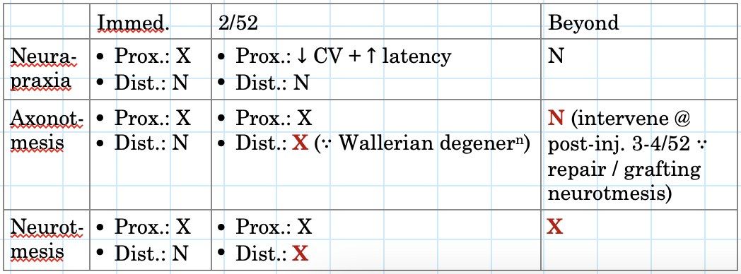
- Neuropraxia: demyelination/myelin sheath intussusception, ischaemia; transient conduction block; complete recovery in days to weeks
- Axonotmesis: axon and myelin sheath degenerate; endo- and perineurium intact; Wallerian degeneration of the distal stump; 30d latency then complete recovery at 1mm/day
- Neurotmesis: complete transection; no recovery unless operated on
Q7What are the effects of graded compression on a nerve?▸
- 0-30mmHg: normal
- 30-50mmHg: decreased epidural venous flow, endoneural oedema, decrease axonal transport
- 50-80mmHg: decreased arterial flow -> no axonal transport
- >80mmHg: complete ischemia
Q8What are the NCS and EMG findings in neuropraxia?▸
- NCS: conduction slowing or conduction block across the level of the lesion but normal distally
- EMG completely stunned state: electrical silence after 3 weeks, no fibrillation as the muscle fibre is not denervated
- EMG partially stunned state: single or limited motor units at high rates
Q9What are the NCS and EMG findings in axonotmesis?▸
- NCS: reduced amplitudes of sensory and motor fibres
- Relative preservation of conduction velocities
- Distal conduction block at around 2 weeks when Wallerian degeneration kicks in
- EMG: positive sharp waves and fibrillations, reduced interference pattern (reduced recruitment)
Q10What are the NCS and EMG findings in neurotmesis?▸
- Initial preservation of distal responses: 3-5 days motor, 6-10 days sensory, then absent sensory and motor responses
- EMG: immediate and complete lack of voluntary activity
- Fibrillation around 2 weeks in UL and 3 weeks in LL - differentiate from severe neuropraxia where there wont be any fibrillations
- Fibrillations abundant and large amplitude in the first 6 months, then diminish as muscle atrophies or fibroses
Q11What are F waves and H reflexes?▸
- F wave: antidromic impulse to AHC then orthodromic conduction down the motor nerve (like an echo); detects proximal lesion; increased F latency with normal motor latency = plexus/root lesion; limited in multi-root injuries
- H reflex: deep tendon reflex - A alpha submaximal stimulation on muscle stretch -> impulse to DRG, monosynaptic reflex via motor neuron, late CMAP; negative in polyneuropathy/radiculopathy; absent in >60yo
- Conditions where no abnormality is seen: pure sensory radiculopathy, before Wallerian degeneration
Q12What does a nerve conduction study consist of?▸
- CMAP (compound muscle action potential)
- SNAP (sensory nerve action potential)
- H reflex
- F wave
Q13In a motor study, what do latency and amplitude reflect?▸
- Latency = quality of axon
- Amplitude = quantity of axon
- Also measure conduction velocity
- Measure distances to calculate the conduction velocity
Q14Describe the technique of performing an NCS.▸
- Room temperature and humidity
- Patient position; appropriate machine settings
- Electrode placement: ortho- or antidromic; ground, active recording, reference and stimulating electrodes (cathode to cathode)
- Stimulate until a satisfactory response; supramaximal stimulation to activate all nerve fibres
- Measure distances to calculate conduction velocity
Q15Describe the degenerative phase of Wallerian degeneration.▸
- Cell body changes from neurotransmission to regeneration: dendrite retracted, chromatolysis, nucleus migrates to periphery, increase RNA production
- Distal to injury: axons and myelin degenerate in an anterograde fashion; Phagocytosis of debris by macrophage and Schwann cells
- Proximal to injury: axon degenerates to the next node of Ranvier
- Occurs in Sunderland II or above
Q16Describe the reparative phase of Wallerian degeneration.▸
- Distal to injury: Schwann cells proliferate forming bridging tubes (bands of Bungner)
- Proximal to injury: axon sprouts (rate 1mm/day) try to grow into the new tube formed by Schwann cells
- Guided by neurotrophic (growth factor) and neurotropic (end organ guidance) factors
- Filopodia = contact guidance
- End organ permanent change if delayed healing (>2 years, 3 months in motor endplate)
Q17What are the prognostic factors for nerve injury recovery?▸
- Patient: age, systemic factors (DM, alcoholism)
- Injury: type of nerve (mixed motor and sensory worse), type and mechanism of injury, distal location better, large gap (>2mm)
- Surgery: timing (delayed repair worse), repair under tension worse, quality of repair
Q18How does a neuroma present and how is it managed?▸
- Clinical: Tinel positive at previous injury site or scar +/- distal atrophy
- In continuity: surgery for local compression, pain, distal denervation; if on-table stimulation negative -> excision and reconstruction
- Terminal: indication is pain -> excision
Q19What is the critical gap for nerve regeneration?▸
- Critical gap 2mm
- A larger gap prevents the regenerating axon from bridging to the distal stump
Q20What are the principles of an ideal nerve repair?▸
- Tension free
- No gap
- End to end with correct orientation of fascicles
- No fascicle extrusion
Q21What are the types of nerve repair?▸
- Epineural: outer connective tissue layer only
- Grouped fascicular repair
- Interfascicular (out of favour)
- Clinically fascicular repair is not superior to epineural repair
Q22How is tension reduced during nerve repair?▸
- Local: mobilisation, transposition
- External: bone shortening
Q23What are the aims and types of nerve grafting?▸
- Aim: provide a scaffold to guide axons to the distal stump
- Nerve graft: autograft or allograft from LABCN, MABCN, AIN, sural or finger (less critical side)
- Nerve conduit: vein graft, silicone tube