24 slides
▸ Slide 32 · Soft tissueSoft Tissue · 2 questions expand
Slide render

Question list
Q1-Q22 questions — tap to reveal all answerslist
- What soft tissue topic is shown on this slide?
- What structures or layers are demonstrated in the slide image?
Answers · Q & A
Q1.What soft tissue topic is shown on this slide?
- Not covered in the speaker notes - the slide image is the only source
Q2.What structures or layers are demonstrated in the slide image?
- Not covered in the speaker notes - the slide image is the only source
▸ Slide 33 · TendonSoft Tissue · 4 questions expand
Slide render
Question list
Q1-Q44 questions — tap to reveal all answerslist
- Outline the hierarchical organisation of tendon, from procollagen to whole tendon.
- What is the collagen building unit within a tendon?
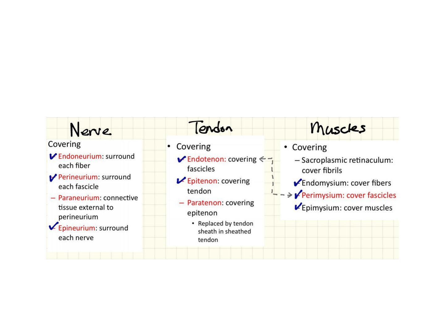
- What connective tissue sheaths surround the subunits of tendon and muscle?
- Outline the hierarchical structure of muscle from sarcomere to whole muscle.
Answers · Q & A
Q1.Outline the hierarchical organisation of tendon, from procollagen to whole tendon.
- Procollagen from cell (1.5nm) -> tropocollagen (280nm) processed by EC protein
- Microfibril: many tropocollagen arranged in a quarter-staggered array, held by cross-links (collagen 11)
- Fibril: multiple microfibrils
- Fibre: multiple fibrils
- 5. fascicle(endotenon)=multiple fiber
- Tendon (epitenon)
Q2.What is the collagen building unit within a tendon?
- 2A1 + 1A2 = collagen from the cell (procollagen, 1.5nm)
- Processed by EC protein to tropocollagen 280nm
- Assembled in a quarter-staggered array held by cross-links
Q3.What connective tissue sheaths surround the subunits of tendon and muscle?
- Tendon: fascicle covered by endotenon, whole tendon by epitenon
- Muscle: fibre covered by endomysium, fascicle by perimysium
Q4.Outline the hierarchical structure of muscle from sarcomere to whole muscle.
- Sarcomere -> myofibril -> fibre (endomysium) -> fascicle (perimysium) -> bundles of fascicles -> muscle
▸ Slide 34 · Nerve cross section and nerve injurySoft Tissue · 5 questions expand
Slide render

Question list
Q1-Q55 questions — tap to reveal all answerslist
- Describe the connective tissue layers of a nerve.
- What is the Erlanger classification of nerve fibres?
- What do the A fibre subtypes supply?
- Describe the sequence of compression damage, recovery and local anaesthetic blocking.
- Which nerve fibre types are myelinated?
Answers · Q & A
Q1.Describe the connective tissue layers of a nerve.
- Neuron -> fibre (endoneurium) -> fascicle (perineurium) -> nerve (epineurium)
Q2.What is the Erlanger classification of nerve fibres?
- A (10-20uM diameter): heavy myelination, fastest conduction
- B (1-3uM): preganglionic autotomic (autonomic); A and B are myelinated
- C (0.5-2um): unmyelinated, slow conduction; postganglionic autonomic, slow pain, thermoreceptors (IV)
- Numerical I, II, III, IV classification is sensory only
Q3.What do the A fibre subtypes supply?
- Aalpha: efferent to skeletal muscle; afferent from muscle spindles (Ia) and tendon stretch organelles (Ib)
- Abeta: organised sensory receptors - Merkel, Meissner, Pacinian, Ruffini, hair follicles (II)
- Agamma: motor to muscle spindle
- Adelta: fast pain (knife), cold sensation, touch (III)
Q4.Describe the sequence of compression damage, recovery and local anaesthetic blocking.
- Sequence of compression damage: A -> C
- Sequence of recovery: C -> A
- Sequence of LA blocking: small myelinated (fast pain) -> unmyelinated -> large myelinated
- Remember to draw the vasa nervorum
Q5.Which nerve fibre types are myelinated?
- A and B fibres are myelinated
- C fibres are unmyelinated (0.5-2um), giving slow conduction
▸ Slide 35 · Seddon classificationSoft Tissue · 9 questions expand
Question list
Q1-Q99 questions — tap to reveal all answerslist
- Describe the Seddon classification of nerve injury.
- What are the effects of graded compression on a nerve?
- What are the NCS and EMG findings in neuropraxia?
- What are the NCS and EMG findings in axonotmesis?
- What are the NCS and EMG findings in neurotmesis?
- What are F waves and H reflexes?
- What does a nerve conduction study consist of?
- In a motor study, what do latency and amplitude reflect?
- Describe the technique of performing an NCS.
Answers · Q & A
Q1.Describe the Seddon classification of nerve injury.
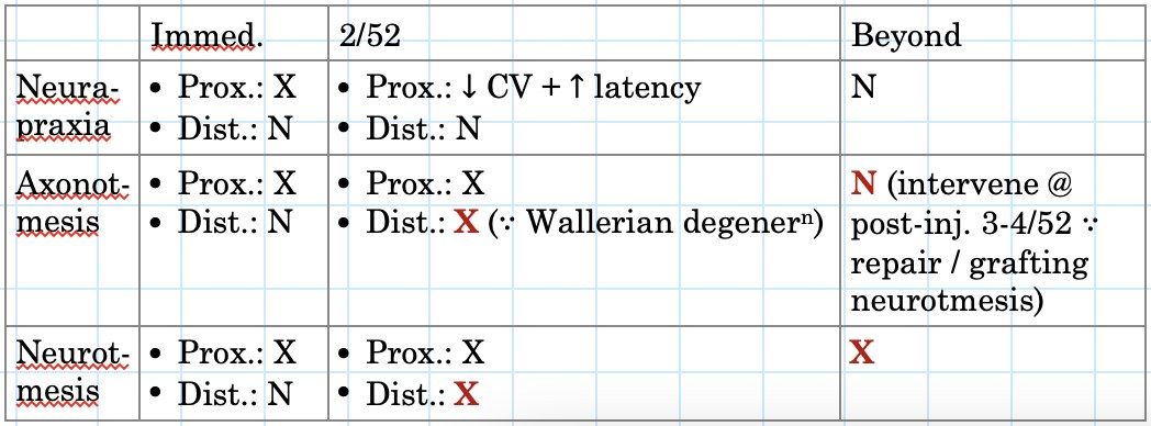
- Neuropraxia: demyelination/myelin sheath intussusception, ischaemia; transient conduction block; complete recovery in days to weeks
- Axonotmesis: axon and myelin sheath degenerate; endo- and perineurium intact; Wallerian degeneration of the distal stump; 30d latency then complete recovery at 1mm/day
- Neurotmesis: complete transection; no recovery unless operated on
Q2.What are the effects of graded compression on a nerve?
- 0-30mmHg: normal
- 30-50mmHg: decreased epidural venous flow, endoneural oedema, decrease axonal transport
- 50-80mmHg: decreased arterial flow -> no axonal transport
- >80mmHg: complete ischemia
Q3.What are the NCS and EMG findings in neuropraxia?
- NCS: conduction slowing or conduction block across the level of the lesion but normal distally
- EMG completely stunned state: electrical silence after 3 weeks, no fibrillation as the muscle fibre is not denervated
- EMG partially stunned state: single or limited motor units at high rates
Q4.What are the NCS and EMG findings in axonotmesis?
- NCS: reduced amplitudes of sensory and motor fibres
- Relative preservation of conduction velocities
- Distal conduction block at around 2 weeks when Wallerian degeneration kicks in
- EMG: positive sharp waves and fibrillations, reduced interference pattern (reduced recruitment)
Q5.What are the NCS and EMG findings in neurotmesis?
- Initial preservation of distal responses: 3-5 days motor, 6-10 days sensory, then absent sensory and motor responses
- EMG: immediate and complete lack of voluntary activity
- Fibrillation around 2 weeks in UL and 3 weeks in LL - differentiate from severe neuropraxia where there wont be any fibrillations
- Fibrillations abundant and large amplitude in the first 6 months, then diminish as muscle atrophies or fibroses
Q6.What are F waves and H reflexes?
- F wave: antidromic impulse to AHC then orthodromic conduction down the motor nerve (like an echo); detects proximal lesion; increased F latency with normal motor latency = plexus/root lesion; limited in multi-root injuries
- H reflex: deep tendon reflex - A alpha submaximal stimulation on muscle stretch -> impulse to DRG, monosynaptic reflex via motor neuron, late CMAP; negative in polyneuropathy/radiculopathy; absent in >60yo
- Conditions where no abnormality is seen: pure sensory radiculopathy, before Wallerian degeneration
Q7.What does a nerve conduction study consist of?
- CMAP (compound muscle action potential)
- SNAP (sensory nerve action potential)
- H reflex
- F wave
Q8.In a motor study, what do latency and amplitude reflect?
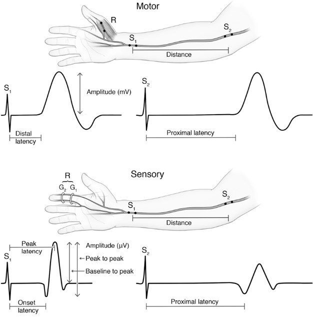
- Latency = quality of axon
- Amplitude = quantity of axon
- Also measure conduction velocity
- Measure distances to calculate the conduction velocity
Q9.Describe the technique of performing an NCS.
- Room temperature and humidity
- Patient position; appropriate machine settings
- Electrode placement: ortho- or antidromic; ground, active recording, reference and stimulating electrodes (cathode to cathode)
- Stimulate until a satisfactory response; supramaximal stimulation to activate all nerve fibres
- Measure distances to calculate conduction velocity
▸ Slide 36Soft Tissue · 2 questions expand

Question list
Q1-Q22 questions — tap to reveal all answerslist
- Which soft tissue structure is the focus of this slide?
- What clinical message does this slide convey?
Answers · Q & A
Q1.Which soft tissue structure is the focus of this slide?
- Not covered in the speaker notes - the slide image is the only source
Q2.What clinical message does this slide convey?
- Not covered in the speaker notes - the slide image is the only source
▸ Slide 37 · EMGSoft Tissue · 6 questions expand
Slide render

Question list
Q1-Q66 questions — tap to reveal all answerslist
- What is EMG and what is its aim?
- Describe the EMG procedure.
- What is measured on EMG and how do neuropathy and myopathy differ?
- What are the EMG findings after nerve injury (axonotmesis/neurotmesis)?
- Compare EMG findings of denervation and myopathy.
- What is a normal interference pattern on EMG?
Answers · Q & A
Q1.What is EMG and what is its aim?
- Studies the electrical activity of individual muscle fibres and motor units
- Aim: differentiate a nerve or muscle problem
- Assess reinnervation
Q2.Describe the EMG procedure.
- Intramuscular needle over different spots for resting and insertional activity
- Voluntary contraction for MUAP
- Maximal voluntary contraction for interference pattern
Q3.What is measured on EMG and how do neuropathy and myopathy differ?
- Insertional activity: increase in neuropathy, decrease in myopathy
- Spontaneous activity (fibrillation, positive sharp wave): increased in neuropathy, decreased in myopathy
- Amplitude: normal in neuropathy, reduced in myopathy
- Recruitment: reduced in neuropathy, increased in myopathy
- Interference pattern: neuropathy reduced with fibrillation; myopathy full but low amplitude
- Duration: increased in neuropathy, decreased in myopathy
Q4.What are the EMG findings after nerve injury (axonotmesis/neurotmesis)?
- Immediately after section the EMG in supplied muscle is normal
- 5-14 days: positive sharp waves consistent with denervation
- 15-30 days: denervation, fibrillation potentials present
- Evidence of re-innervation: highly polyphasic motor unit potentials
Q5.Compare EMG findings of denervation and myopathy.
- Resting: both have fibrillation
- Slight contraction: denervation giant unit; myopathy small polyphasic unit
- Maximal contraction: denervation reduced interference pattern; myopathy full pattern but decreased magnitude
Q6.What is a normal interference pattern on EMG?
- A disorderly group of action potentials of varying rates and amplitudes
▸ Slide 38 · Draw a wave form of NCSSoft Tissue · 2 questions expand
Question list
Q1-Q22 questions — tap to reveal all answerslist
- Describe how you would draw the waveform of a nerve conduction study.
- What are the components of an NCS waveform?
Answers · Q & A
Q1.Describe how you would draw the waveform of a nerve conduction study.
- Not covered in the speaker notes - the slide only asks to draw the NCS waveform
Q2.What are the components of an NCS waveform?
- Not covered in the speaker notes
▸ Slide 39 · Wallerian degenerationSoft Tissue · 5 questions expand
Slide render

Question list
Q1-Q55 questions — tap to reveal all answerslist
- Describe the degenerative phase of Wallerian degeneration.
- Describe the reparative phase of Wallerian degeneration.
- What are the prognostic factors for nerve injury recovery?
- How does a neuroma present and how is it managed?
- What is the critical gap for nerve regeneration?
Answers · Q & A
Q1.Describe the degenerative phase of Wallerian degeneration.
- Cell body changes from neurotransmission to regeneration: dendrite retracted, chromatolysis, nucleus migrates to periphery, increase RNA production
- Distal to injury: axons and myelin degenerate in an anterograde fashion; Phagocytosis of debris by macrophage and Schwann cells
- Proximal to injury: axon degenerates to the next node of Ranvier
- Occurs in Sunderland II or above
Q2.Describe the reparative phase of Wallerian degeneration.
- Distal to injury: Schwann cells proliferate forming bridging tubes (bands of Bungner)
- Proximal to injury: axon sprouts (rate 1mm/day) try to grow into the new tube formed by Schwann cells
- Guided by neurotrophic (growth factor) and neurotropic (end organ guidance) factors
- Filopodia = contact guidance
- End organ permanent change if delayed healing (>2 years, 3 months in motor endplate)
Q3.What are the prognostic factors for nerve injury recovery?
- Patient: age, systemic factors (DM, alcoholism)
- Injury: type of nerve (mixed motor and sensory worse), type and mechanism of injury, distal location better, large gap (>2mm)
- Surgery: timing (delayed repair worse), repair under tension worse, quality of repair
Q4.How does a neuroma present and how is it managed?
- Clinical: Tinel positive at previous injury site or scar +/- distal atrophy
- In continuity: surgery for local compression, pain, distal denervation; if on-table stimulation negative -> excision and reconstruction
- Terminal: indication is pain -> excision
Q5.What is the critical gap for nerve regeneration?
- Critical gap 2mm
- A larger gap prevents the regenerating axon from bridging to the distal stump
▸ Slide 40 · Nerve repair and graftingSoft Tissue · 4 questions expand
Slide render
Question list
Q1-Q44 questions — tap to reveal all answerslist
- What are the principles of an ideal nerve repair?
- What are the types of nerve repair?
- How is tension reduced during nerve repair?
- What are the aims and types of nerve grafting?
Answers · Q & A
Q1.What are the principles of an ideal nerve repair?
- Tension free
- No gap
- End to end with correct orientation of fascicles
- No fascicle extrusion
Q2.What are the types of nerve repair?
- Epineural: outer connective tissue layer only
- Grouped fascicular repair
- Interfascicular (out of favour)
- Clinically fascicular repair is not superior to epineural repair
Q3.How is tension reduced during nerve repair?
- Local: mobilisation, transposition
- External: bone shortening
Q4.What are the aims and types of nerve grafting?
- Aim: provide a scaffold to guide axons to the distal stump
- Nerve graft: autograft or allograft from LABCN, MABCN, AIN, sural or finger (less critical side)
- Nerve conduit: vein graft, silicone tube
▸ Slide 41 · Resting potentialSoft Tissue · 4 questions expand
Slide render

Question list
Q1-Q44 questions — tap to reveal all answerslist
- What is the resting membrane potential and ionic distribution?
- What maintains the resting potential?
- Describe the action potential.
- What determines conduction velocity and how do local anaesthetics work?
Answers · Q & A
Q1.What is the resting membrane potential and ionic distribution?
- Resting potential -70mV
- Intracellular: high K, organic ions
- Extracellular: high Na, high Cl-
Q2.What maintains the resting potential?
- Lipid membrane with selective permeability to ions (more permeable to K)
- Active Na/K pump (needs ATP)
- Donnan equilibrium - impermeable proteins alter distribution of permeable ions to restore osmotic equilibrium
Q3.Describe the action potential.
- Threshold stimulus (can be summation of smaller stimuli)
- Na channels open, Na influx -> depolarisation (-70mV to +30mV); close spontaneously after 1ms
- K channels open, K efflux -> repolarisation (below -70mV)
- Na/K exchange pump restores resting potential
- Refractory period from Na inactivation and K hyperpolarisation ensures unilateral propagation
- Myelinated nerves propagate at nodes of Ranvier (saltatory conduction)
Q4.What determines conduction velocity and how do local anaesthetics work?
- Conduction velocity depends on the type of nerve fibre and myelination
- Local anaesthetics work at Na channels, blocking propagation and initiation of the AP by plugging the Na channels
▸ Slide 42 · Action potential arrive NMJSoft Tissue · 5 questions 1 check expand
Slide render
Question list
Q1-Q55 questions — tap to reveal all answerslist
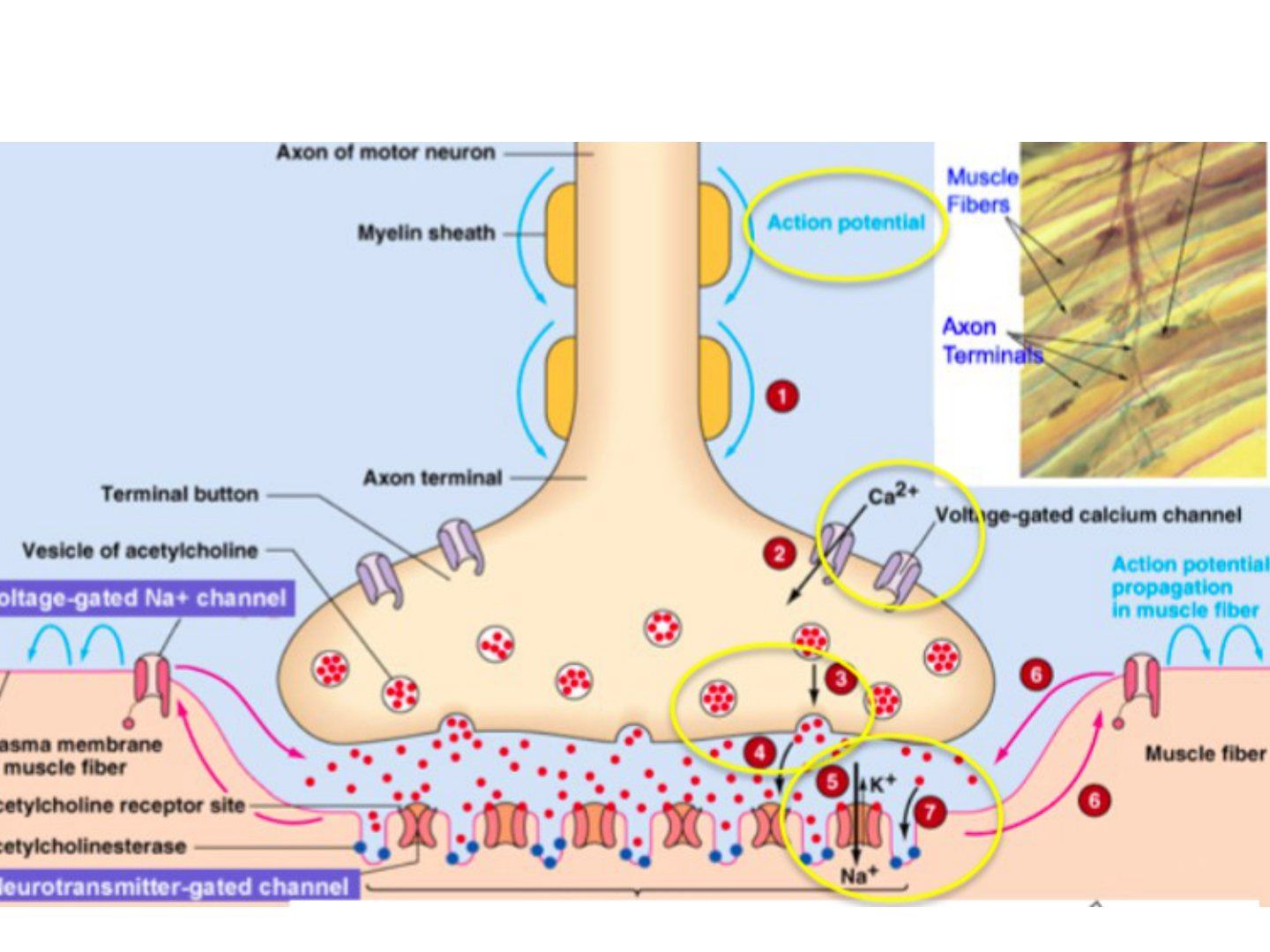
- Describe the events when an action potential arrives at the NMJ.
- How does Botox act at the NMJ?
- Compare depolarising and non-depolarising neuromuscular blockers.
- What is the pathophysiology of myasthenia gravis?
- Which other toxin acts at the neuromuscular junction?
Answers · Q & A
Q1.Describe the events when an action potential arrives at the NMJ.
- Resting potential changes from -70mV to +30mV
- Ca influx into neuron via voltage-gated calcium channels
- Release of Ach
- Ach binds postsynaptic nicotinic sodium channels
- Increased Na influx -> AP propagates along the sarcolemma and down the T tubules
- Ca release from the sarcoplasmic reticulum; Ca binds troponin C, moves tropomyosin, exposing the myosin binding site on actin
Q2.How does Botox act at the NMJ?
- inhibit SNAP 25 protein
- SNAP 25 is important in production of Ach vesicles and migration of the vesicles to the junction
- Therefore prevents release of Ach
Q3.Compare depolarising and non-depolarising neuromuscular blockers.
- Depolarising - succinylcholine: hyperpolarises the sarcolemma (per speaker notes)
- Non-depolarising - rocuronium: prevents depolarisation of the sarcolemma
Q4.What is the pathophysiology of myasthenia gravis?
- Antibodies against the receptor
Q5.Which other toxin acts at the neuromuscular junction?
- Tetanus (toxin) is also mentioned alongside Botox as acting at the NMJ
Fact check
Depolarising NM blockade with succinylcholine hyperpolarises the sarcolemma — error - mechanism wrong — succinylcholine is an ACh receptor agonist causing persistent depolarisation of the motor endplate (inactivating sodium channels), not hyperpolarisation — source
▸ Slide 43 · MuscleSoft Tissue · 8 questions expand

Question list
Q1-Q88 questions — tap to reveal all answerslist
- Starting from the sarcomere, list the levels of organisation of a skeletal muscle.
- Classify muscles by shape and fascicular structure.
- What are the types of muscle contraction?
- Compare open and closed chain exercises.
- Compare type 1 and type 2 muscle fibres.
- What is the response of muscle to injury?
- How do skeletal, cardiac and smooth muscle differ in nuclei?
- According to which criteria are muscles classified?
Answers · Q & A
Q1.Starting from the sarcomere, list the levels of organisation of a skeletal muscle.
- Sacromere > myofibril > muscle fibre (myofibril + SR, endomysium) > muscle fascicle (perimysium) > single muscle (epimysium)
- At the musculotendinous junction fibres run from tendon endotenon to muscle perimysium
- Muscle involvement increases surface area to distribute load transfer
Q2.Classify muscles by shape and fascicular structure.
- Parallel: more ROM due to length of fibres, but less total force due to less physiological cross sectional area - strap (sartorius), fan (pec major), fusiform (biceps)
- Pennate: more fibres per cross-sectional area, more power but less ROM - unipennate (EDL), bipennate (rectus femoris), multipennate (deltoid)
- The horizontal component makes the pennate muscle belly solid and compact
Q3.What are the types of muscle contraction?
- Isometric: force generated but no change in length; adv co-contraction with no shearing, protects joint; disadv limited performance gain
- Isotonic: constant tension with length change - concentric shorten, eccentric lengthen; adv allows joint motion; disadv speed not well controlled, resistance depends on gravity
- Isokinetic: speed constant through full ROM while load changes to maintain velocity; adv maximises strength
Q4.Compare open and closed chain exercises.
- Closed chain: most distal segment stabilised and not moving; allows muscle co-contraction, generates compression force, more functional
- Open chain: extremity free to move; generates shear force; for training a particular muscle/muscle group
Q5.Compare type 1 and type 2 muscle fibres.
- Type 1: red, aerobic (Kreb cycle - glycogen + fatty acids -> oxidative phosphorylation -> ATP), less strong, slower, endurance, first to atrophy in deconditioning [slow red ox]
- Type 2: white, anaerobic (ATP-CP, glycolysis producing lactic acid), stronger, fast acting
Q6.What is the response of muscle to injury?
- Mild: DOMS - oedema and inflammation after unaccustomed eccentric exercise (24-48hrs, increased CK); strain - inflammation at MTJ from eccentric contraction, complication fibrosis
- Atrophy: disuse/neuropathy, affects monoarticular more than biarticular, increased fatigability, fatty infiltration on histology
- Laceration: limited recovery; distal stump denervation -> atrophy -> soft tissue bridge between tendon and muscle belly
- Repair phases: inflammatory (1/7), proliferative (10/7) (satellite cell -> myoblast, fibrin -> scar), remodel (90 days)
- Repair needs blood supply and innervation
Q7.How do skeletal, cardiac and smooth muscle differ in nuclei?
- Skeletal muscle: multinucleated
- Cardiac: single central nucleus
- Smooth muscle: single central nucleus
Q8.According to which criteria are muscles classified?
- Shape and fascicular structure
- Myoglobin content
- Bi- or mono-articular
- Orientation of line of pull relative to the joint surface (flexor, extensor, adductor, abductor)
- Type of contractile activity
▸ Slide 44 · SarcomereSoft Tissue · 4 questions expand
Slide render
Question list
Q1-Q44 questions — tap to reveal all answerslist
- Describe the bands of the sarcomere.
- Describe the structure of actin and myosin.
- Describe the Huxley sliding theory of muscle contraction.
- Where does the ATP for contraction come from?
Answers · Q & A
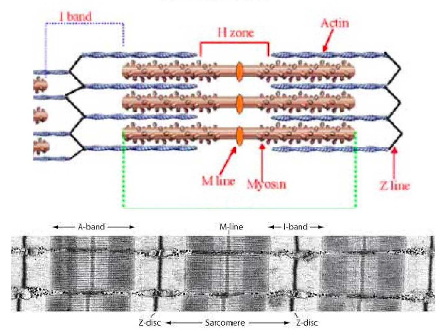
Q1.Describe the bands of the sarcomere.
- A band: where actin and myosin overlap (double refractive)
- I band (light): just actin (single refractive)
- H zone: just myosin
- H and I bands decrease with contraction
- M line anchors myosin in a hexagonal lattice (1M=6A)
- Z line anchors actin in a tetragonal lattice
Q2.Describe the structure of actin and myosin.
- Actin: two chains of beads in a double helix
- Myosin: golf clubs with two heads
Q3.Describe the Huxley sliding theory of muscle contraction.
- Tropomyosin surrounds actin
- Ca influx from the SR after AP arrival at the T tubules binds troponin C (I inhibitory, T binds troponin to tropomyosin), causing conformational change of troponin-tropomyosin complex
- Exposing the myosin binding site; myosin head attaches forming a cross bridge
- ATP hydrolysis causes S1 myosin head rotation -> power stroke; filaments slide (I and H band will decrease)
- ATP binding will break cross bridge and reset myosin heads
Q4.Where does the ATP for contraction come from?
- Kreb cycle - aerobic: glycogen + fatty acids -> oxidative phosphorylation -> ATP
- ATP-CP - anaerobic, glycolysis which produces lactic acid
▸ Slide 45 · Optimal length =Soft Tissue · 4 questions expand
Slide render
Question list
Q1-Q44 questions — tap to reveal all answerslist
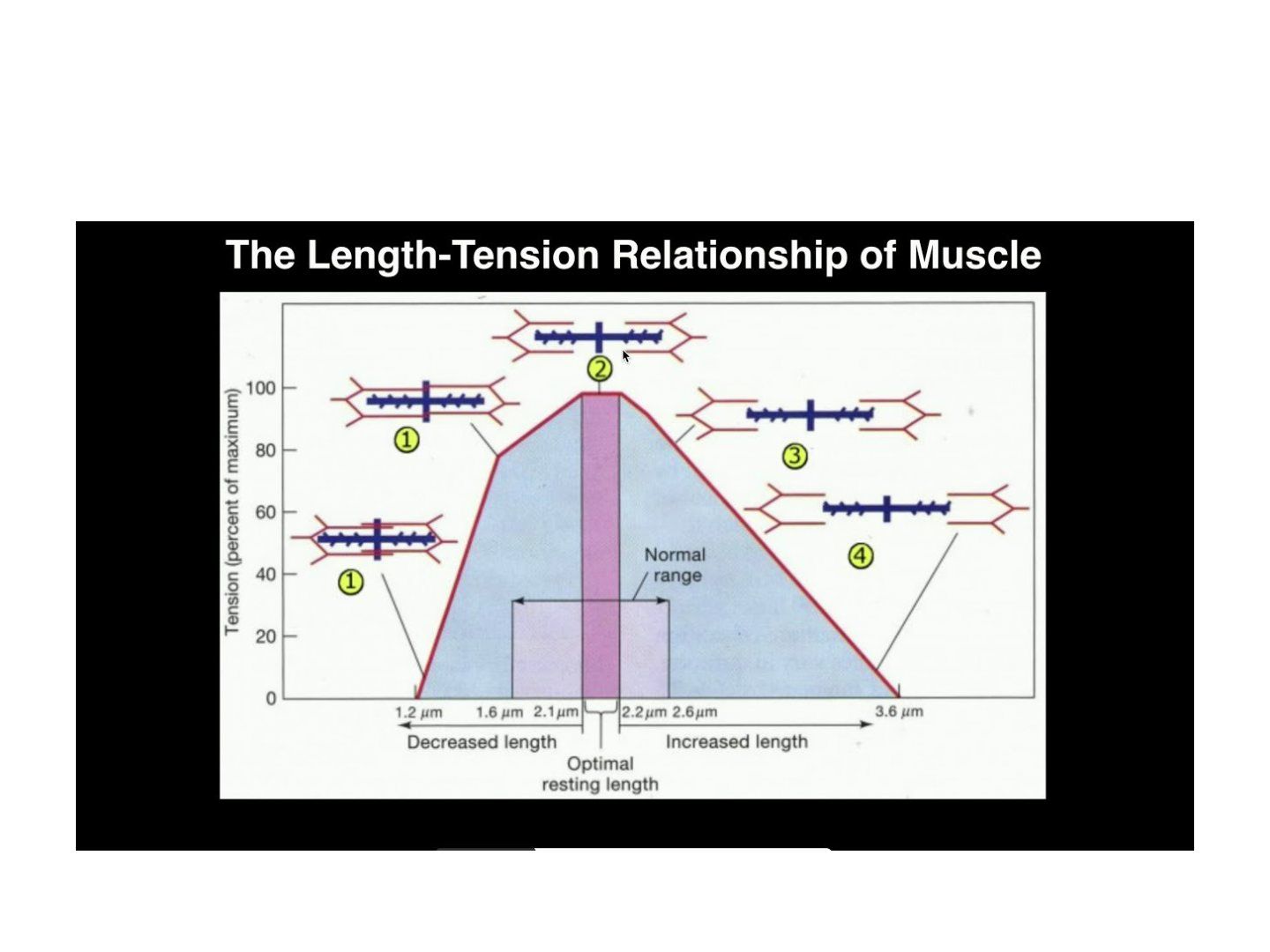
- What is meant by optimal length of a muscle?
- What is the working range of muscle around optimal length?
- Give the clinical example of muscle working length given in the lecture.
- Which cardiac law is related to the concept of optimal length?
Answers · Q & A
Q1.What is meant by optimal length of a muscle?
- Length allowing the maximal number of cross bridge formation
- No overlap of thin filaments
Q2.What is the working range of muscle around optimal length?
- 70-130% of optimal length
Q3.Give the clinical example of muscle working length given in the lecture.
- Abductor tension during THR (total hip replacement)
Q4.Which cardiac law is related to the concept of optimal length?
- The Frank-Starling Law
▸ Slide 46 · CollagenSoft Tissue · 8 questions expand
Question list
Q1-Q88 questions — tap to reveal all answerslist
- Describe the synthesis of collagen.
- What is the collagen chain sequence shown on the slide and which structures does it form?
- What proportion of whole body proteins is collagen?
- What are the features of type I collagen?
- Where is type II collagen found?
- Where is type III collagen found and what is its role?
- Where is type IV collagen found and with what is it associated?
- Where are collagen types V, VI, XI and X found?
Answers · Q & A
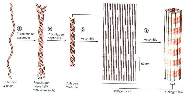
Q1.Describe the synthesis of collagen.
- Amino acids such as glycine-proline-X or glycine-X-hydroxyproline form procollagen polypeptide chains in the endoplasmic reticulum
- Hydroxylation and glycosylation allow three polypeptide chains to form a right-handed triple superhelix (two alpha-1 and one alpha-2 chain)
- Cleaved extracellularly to become tropocollagen, then arranged into quarter-staggered arrays by cleavage of the N and C terminal propeptidases -> collagen microfibrils
- Aggregation of collagen microfibrils by formation of covalent crosslinks by lysyl oxidase -> collagen fibres
Q2.What is the collagen chain sequence shown on the slide and which structures does it form?
- Gly-X-Y-Gly-X-Y-Gly-X-Y
- -> procollagen polypeptide chains (e.g. pro alpha chains) -> procollagen (triple helix) -> collagen microfibril -> collagen fibre
Q3.What proportion of whole body proteins is collagen?
- 25% of whole body proteins
Q4.What are the features of type I collagen?
- 90% of total collagen
- Bone, tendon, ligament and skin
- Annular fibrosis
- Associated with OI (osteogenesis imperfecta) and EDS (Ehlers-Danlos syndrome)
Q5.Where is type II collagen found?
- Hyaline cartilage
- Nucleus pulposus
Q6.Where is type III collagen found and what is its role?
- Damage and repair collagen
- Skin and blood vessels
- Dupuytren contracture and frozen shoulder
Q7.Where is type IV collagen found and with what is it associated?
- Basement membrane (BM)
- Nephrotic syndrome
Q8.Where are collagen types V, VI, XI and X found?
- V, VI, XI - cartilage (articular surface) and MED (multiple epiphyseal dysplasia)
- X - cartilage (deep calcified layer) and early OA
▸ Slide 47 · What is cartilage?Soft Tissue · 17 questions expand

Question list
Q1-Q1717 questions — tap to reveal all answerslist
- What are the features of articular cartilage?
- List the types of cartilage with examples.
- What is the composition of articular cartilage?
- What are the biomechanical properties of cartilage and what explains the biphasic nature?
- Describe the zones of articular cartilage.
- What is the lamina splendens?
- How are load and stiffness transitioned from cartilage to bone?
- How does cartilage retain water and what happens in OA?
- What is the structure of a proteoglycan?
- What are the functions of proteoglycans in cartilage?
- Describe the microscopic changes of osteoarthritis.
- Describe the macroscopic changes of osteoarthritis.
- What are the changes of cartilage aging (as opposed to OA)?
- What are the X-ray findings of osteoarthritis?
- How does articular cartilage respond to injury?
- What is the function of matrix glycoproteins?
- What are the matrix regions of cartilage?
Answers · Q & A
Q1.What are the features of articular cartilage?
- Avascular, aneural, alymphatic, non-immunogenic
- Decreases friction (coefficient 0.002) - 3x more slippery than ice on ice
- Distributes load - resists tensile, shear and compressive forces
- Poor intrinsic healing capacity; nutrition via diffusion from synovial fluid
- Maintains joint congruence and absorbs shock
Q2.List the types of cartilage with examples.
- Hyaline - nasal, trachea
- Fibrocartilage - ACJ, SCJ, IVD, labrum, meniscus (less water, fewer cells, more matrix)
- Elastic - ligamentum flavum, epiglottis
- Hyaline is the most common and forms articular surfaces, costal cartilages and growth plates
- Cartilage type reflects function: fibrocartilage resists tension, elastic maintains shape (e.g. ear, epiglottis)
Q3.What is the composition of articular cartilage?
- Cells 5% (chondrocytes) - synthesise matrix
- Water 75%; ECM 20%
- Collagen 10% (type II in hyaline) - resists tensile forces
- Proteoglycan 10% - resists compressive force
- Matrix glycoproteins (fibronectin, chondronectin), degradative enzymes
- Solid matrix = collagen + proteoglycan; fluid phase = water + dissolved ions
Q4.What are the biomechanical properties of cartilage and what explains the biphasic nature?
- Anisotropic, biphasic, viscoelastic
- Biphasic = water-soaked sponge: solid collagen-proteoglycan phase plus fluid phase (permeability)
- Under high compression, water movement is hindered by frictional drag, reduced porosity and negative PG charges, giving greater stiffness
- Under tension, the crosslinks of the collagen fibres are pulled apart, increasing the water permeability, which leads to decrease in stiffness
- Viscoelastic = sensitive to the speed and duration of loading
- Creep occurs with sustained load; stress relaxation reduces force under constant deformation
Q5.Describe the zones of articular cartilage.
- Superficial gliding layer 20%: flat chondrocytes (highest number), tangential collagen, highest collagen and water, lowest PG, Water squeezed out to produce squeeze film lubrication
- Middle transition layer 50%: round chondrocytes, oblique fibres at 90 degrees, higher PG
- Deep radial layer 30%: columnar chondrocytes, Collagen fibres are largest diameters, vertically oriented and anchored to subchondral bone, Highest concentration of PG, Lowest H2O content
- Tidemark: smooth, undulating basophilic line for transmission of load between cartilage and bone; cell-free undulating calcification front resisting shear; migrates toward the surface with age
- Calcified zone: collagen X and hydroxyapatite anchor cartilage to subchondral bone with a gradual transition of mechanical properties
Q6.What is the lamina splendens?
- No cells - a thin layer of collagen fibrils with little proteoglycan
- A cellular layer of flattened chondrocytes 1-3 layers thick
Q7.How are load and stiffness transitioned from cartilage to bone?
- Calcified zone anchors the layers using collagen X and hydroxyapatite crystals
- Allows gradual transition of mechanical properties between cartilage and bone
- Subchondral and cancellous bone give a gradual transition of Young's modulus to evenly distribute load
- The fibre architecture is described as the arcades of Benninghoff
Q8.How does cartilage retain water and what happens in OA?
- The negative charge of the proteoglycan creates a repulsion force which is neutralized by the positive ions in the surrounding fluid (i.e. water). The ionic pressure creates swelling pressure which will keep soaking up water until it is resisted by the tension of the collagen fibres
- In OA the collagen network degrades, water content increases, and elasticity/elastic modulus decrease
- Injury beyond the tidemark bleeds and heals with fibrocartilage (type I and II collagen)
- Water content is highest in the superficial zone and decreases with depth; PG concentration shows the opposite gradient
Q9.What is the structure of a proteoglycan?
- Hyaluronate backbone with many GAG side chains, secreted by chondrocytes
- Link proteins connect onto the protein core: G1, G2 near the N terminus (HA backbone) and G3 near the C terminus
- Keratin sulfate (shorter, closer to HA) and chondroitin sulfate joined by sugar bonds
- The whole structure is called an aggregate
Q10.What are the functions of proteoglycans in cartilage?
- Fill the interstices between collagen fibrils
- Electrostatic repulsion maintains tension between fibrils, providing compressive strength
- Traps water due to the hydrophilic aggrecan
Q11.Describe the microscopic changes of osteoarthritis.
- OA (osteoarthritis) is a non-inflammatory joint disease characterized by cartilage loss, new bone formation and capsular fibrosis; Imbalance between repair and degradation
- Abnormal mechanical loading -> chondrocyte damage -> production of proteases, MMP, IL1
- Collagen meshwork breakdown increases permeability
- PG production and degradation both increase but net amount decreases; chains shorten with chondroitin:keratin ratio increased (C4S up, KS down)
- Decreased Young's modulus; stress concentrates on subchondral bone causing subchondral damage
Q12.Describe the macroscopic changes of osteoarthritis.
- Cartilage: softening, fibrillation, fissures or gross erosions
- Bone: subchondral thickening and osteophytes (metaplasia, endochondral ossification via the Indian hedgehog pathway)
- Synovium: synovial inflammation and joint capsule hypertrophy
Q13.What are the changes of cartilage aging (as opposed to OA)?
- Decline in the ability of chondrocytes to maintain matrix: density decreases, cell size increases
- chondrocytes become less responsive to the proliferative and anabolic effects of growth factors, with failure of homoeostasis and inability to withstand external mechanical stresses; PG activity decreases
- Marked increase in advanced glycation end products (AGEs) -> more collagen crosslinking -> stiffer, more susceptible to fatigue failure
Q14.What are the X-ray findings of osteoarthritis?
- Decreased joint space - decreased articular cartilage
- Osteophytes - subchondral bone neovascularisation, proliferation of cartilage and woven bone via endochondral ossification through the Indian hedgehog pathway
- Subchondral cyst - areas of focal bone necrosis
- Subchondral sclerosis
Q15.How does articular cartilage respond to injury?
- Superficial laceration - does not heal
- Deep laceration - heals with fibrocartilage (type I collagen, no organised zones); haemorrhage -> fibrin clot -> growth factors and fibroblasts
- Contusion - chondrocyte death, matrix damage, fissuring, fibrillation and swelling
Q16.What is the function of matrix glycoproteins?
- Interact with collagen fibrils and stabilise the matrix framework
- Help chondrocytes bind to matrix macromolecules (type 6 collagen)
- Act as tissue glue binding the various matrix components
Q17.What are the matrix regions of cartilage?
- Pericellular matrix - plays a role to initiate signal transduction within cartilage with load bearing
- Territorial matrix - protects chondrocytes against mechanical stresses and contributes to resiliency
- Interterritorial matrix - contributes most to the biomechanical properties
- Regions differ in collagen content, fibril diameter/orientation and proteoglycan/non-collagenous protein content
▸ Slide 48 · Difference in type of collagen, chondrocyte number, size of collagen fibres, perSoft Tissue · 2 questions expand
Slide render
Question list
Q1-Q22 questions — tap to reveal all answerslist
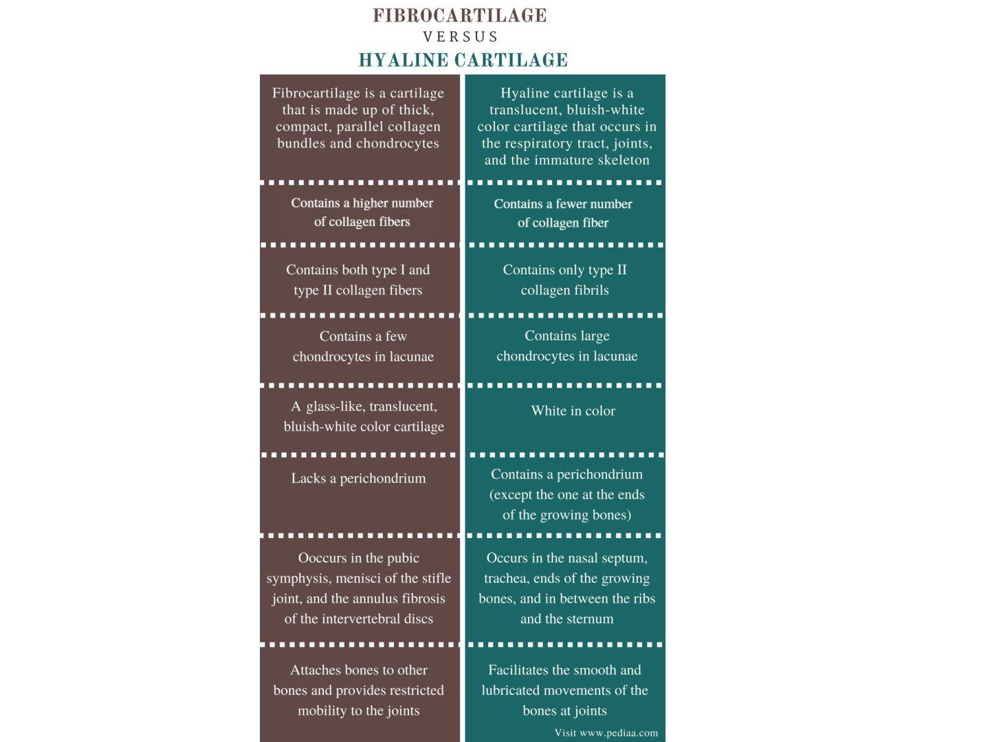
- What physiological stress is applied to cartilage?
- What structural features differ between zones and types of cartilage?
Answers · Q & A
Q1.What physiological stress is applied to cartilage?
- Cyclic loading (1-5 MPa)
- Moderate frequency (<1 Hz)
- Low rate (<1000 MPa/s)
Q2.What structural features differ between zones and types of cartilage?
- Type of collagen
- Chondrocyte number
- Size of collagen fibres
- Presence of perichondrium
▸ Slide 49 · Synovial fluidSoft Tissue · 5 questions expand
Question list
Q1-Q55 questions — tap to reveal all answerslist
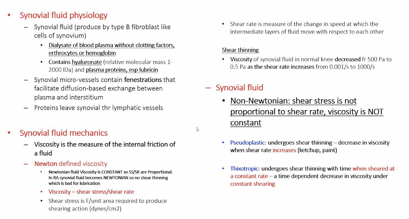
- What are the functions of synovial fluid?
- How is synovial fluid synthesised?
- What are the two synovial cell types?
- What are the main components of synovial fluid?
- What are the rheological properties of synovial fluid?
Answers · Q & A
Q1.What are the functions of synovial fluid?
- Lubrication of the joint
- Cartilage nourishment
Q2.How is synovial fluid synthesised?
- Plasma ultrafiltrate
- Plus secretion from the synovial membrane
Q3.What are the two synovial cell types?
- Type A - macrophage-derived, involved in phagocytosis
- Type B - fibroblast-like, secrete hyaluronate, fibronectin and collagen
Q4.What are the main components of synovial fluid?
- Lubricin - provides lubrication
- Hyaluronan - elastic solid during high strain actions
Q5.What are the rheological properties of synovial fluid?
- Non-Newtonian
- Thixotropic
- Pseudoplastic
▸ Slide 50 · Resting zoneSoft Tissue · 8 questions expand
Slide render

Question list
Q1-Q88 questions — tap to reveal all answerslist
- Describe the resting zone of the growth plate.
- Describe the proliferative zone.
- What happens in the zone of hypertrophy?
- What forms the primary and secondary spongiosa?
- What are the groove of Ranvier and the perichondral ring of Lacroix?
- How is the growth plate regulated and what is its blood supply?
- What did Urist (Science 1965) discover?
- What are the clinical uses of BMPs?
Answers · Q & A
Q1.Describe the resting zone of the growth plate.
- High proportion of ECM to cells, roundish cells
- Cells have high lipid body and vacuole content for later nutritional requirements
- Stem cell characteristics, relatively quiescent with low proliferation
- Low oxygen tension; PTHrP expressing chondrocytes contribute to formation of columnar chondrocytes through the PTHrP –Indian Hedgehog feedback loop)
- Implicated in Gaucher's disease
Q2.Describe the proliferative zone.
- Chondrocytes are flat and arranged in columns
- Matrix production (type II collagen and proteoglycan) and cell division give longitudinal growth
- High oxygen tension, good blood supply
- Implicated in achondroplasia
Q3.What happens in the zone of hypertrophy?
- Zone of maturation: Cell division ceases and chondrocytes increase in size 5X; prepare matrix for calcification
- Zone of degeneration: calcium accumulated in mitochondria is released when glycogen is depleted
- Zone of provisional calcification: calcified chondroid matrix becomes scaffold for bone deposition, high ALP
- Implicated in SCFE, rickets, physeal fracture
Q4.What forms the primary and secondary spongiosa?
- Primary: vascular invagination and woven bone; osteoblasts line up on the calcified cartilage bar (low O2, good blood supply); site of acute osteomyelitis
- Secondary: remodelling - internal (removes cartilage bar and woven bone to lamellar bone), external (funnelisation)
- Implicated in osteopetrosis, osteogenesis imperfecta and scurvy
Q5.What are the groove of Ranvier and the perichondral ring of Lacroix?
- Groove of Ranvier: contains osteoblasts, fibroblasts and chondrocytes; responsible for appositional growth
- Perichondral ring of Lacroix: dense fibrous tissue anchoring epiphysis to metaphysis
Q6.How is the growth plate regulated and what is its blood supply?
- Growth factors (IGF, FGF, TGF, PDGF, TNF), vitamins and hormones
- Epiphysis: epiphyseal artery (only to proliferative zone)
- Physis: perichondral artery predominant
- Metaphysis: metaphyseal artery; diaphysis: nutrient artery
Q7.What did Urist (Science 1965) discover?
- Demineralised bone fragments implanted intramuscularly or subcutaneously induced bone formation
- Led to the discovery of BMP-2 and BMP-7
Q8.What are the clinical uses of BMPs?
- FDA approved for acute tibial fractures and complex nonunions
- BMP-2 is preferred as it is more effective and less costly
▸ Slide 51 · Tell me about the meniscusSoft Tissue · 13 questions expand
Slide render

Question list
Q1-Q1313 questions — tap to reveal all answerslist
- What are the functions of the meniscus?
- How much compressive load does the meniscus take up?
- What is the composition and macrostructure of the meniscus?
- Describe the lateral meniscus.
- Describe the medial meniscus.
- What are the meniscal ligaments?
- What is the blood supply of the meniscus?
- How does the meniscus bear load?
- How does the meniscus absorb shock?
- How does the meniscus provide lubrication and nutrition?
- How does the meniscus stabilise the knee?
- What contact area and stress changes follow partial and total meniscectomy?
- What is the association of SONK with meniscal root tears?
Answers · Q & A
Q1.What are the functions of the meniscus?
- Load transmission - increases contact area, decreases peak stress
- Shock absorption via viscoelasticity
- AP stabilisation
- Lubrication and nutrition
- Proprioception (mechanoreceptors in the meniscal horns)
- Mnemonic SSLLA
Q2.How much compressive load does the meniscus take up?
- 60% of compressive load in extension
- 90% of compressive load at 90 degrees of flexion
Q3.What is the composition and macrostructure of the meniscus?
- Cells: fibrochondrocytes (middle/inner), fibroblast-like cells (outer half), superficial zone cells
- ECM: water 70%, collagen 22% (outer type I, inner type II), proteoglycans, non-collagen proteins
- Superior superficial layer (random fibres); superior lamellar layer (radial fibres)
- Deep layer - fibres run circumferentially to resist hoop stresses
- Inferior lamellar layer (radial and random); inferior superficial layer (radial)
Q4.Describe the lateral meniscus.
- O-shaped, covers 80-85% of the lateral tibial plateau, takes 70% of compartment load
- 40-50% contact area reduced after meniscectomy, 200% increase in stress
- Anterior horn just posterior to the ACL insertion, to which it partially blends; posterior horn anterior to the MM posterior horn
- No attachment to the LCL; loose attachment to the capsule
Q5.Describe the medial meniscus.
- C-shaped, covers 60-65% of the medial plateau, takes 50% of compartment load
- Larger AP diameter than width
- 50-70% contact area reduced after meniscectomy, 100% increase in contact stress
- Anterior root 7 mm anterior to the ACL; posterior root anterior to the PCL
- Attaches to the deep MCL and capsule via the coronary ligament
Q6.What are the meniscal ligaments?
- Insertional ligament (meniscotibial ligament)
- Intermeniscal ligament
- Meniscofemoral ligament
- Attachment to the deep medial collateral ligament
Q7.What is the blood supply of the meniscus?
- Blood supply reaches the periphery from perimeniscal plexus (lateral and medial inferior genicular arteries), supplying the peripheral 10-30%
- Central 2/3 nourished by diffusion
- Posterior horn supplied by the middle geniculate artery
- Zones: red-red, red-white, white-white
Q8.How does the meniscus bear load?
- Compressive axial forces are converted to a radially directed force taken up as circumferential hoop stress
- Radial fibres act as intrasubstance tie-rods resisting longitudinal splitting and excessive compression
- The hoop must be complete - intact circumference and bony attachments
- Tensile modulus: Hoop 110Mpa, Radial 10Mpa
- Barrel analogy: the wedged cross-section extrudes radially, increasing circumference and generating hoop stress
Q9.How does the meniscus absorb shock?
- Intact menisci dissipate force as biphasic structures
- Reduce 20% of peak force on articular cartilage and bone by increasing TFJ conformity
- The collagen-GAG network resists water movement through the solid phase
- Meniscal tissue is less stiff than articular cartilage because of the lower concentration of proteoglycans; Meniscal tissue is more resistant to the internal movement of water through its tissue than articular cartilage
Q10.How does the meniscus provide lubrication and nutrition?
- By increasing joint congruity it allows better fluid entrainment and hydrodynamic lubrication
- The sponge phenomenon helps circulate cellular nutrients throughout the joint
Q11.How does the meniscus stabilise the knee?
- Medial meniscus posterior horn resists anterior tibial translation in an ACL-deficient knee
- Meniscofemoral ligaments are secondary restraints to posterior drawer
- The meniscus construct is a restraint to tibial rotation
Q12.What contact area and stress changes follow partial and total meniscectomy?
- Partial meniscectomy of the inner third: contact area -10%, peak load +65%
- Total meniscectomy: contact area -75%, peak load +235%
Q13.What is the association of SONK with meniscal root tears?
- SONK (spontaneous osteonecrosis of the knee) has been suggested to relate to root tears
- Female:male ratio 3:1
▸ Slide 52 · Intervertebral discSoft Tissue · 8 questions expand
Question list
Q1-Q88 questions — tap to reveal all answerslist
- What are the functions of the spine and intervertebral disc?
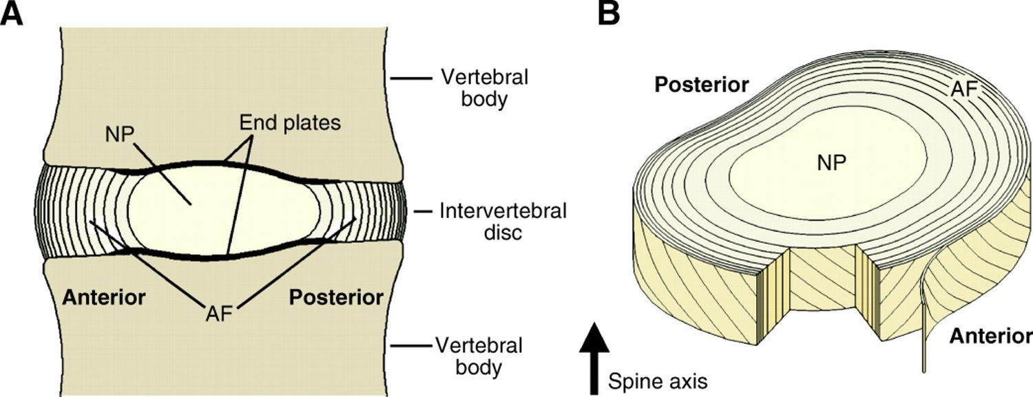
- What are the components of the intervertebral disc?
- Describe the structure of the annulus fibrosus.
- What is the blood and nerve supply of the disc?
- What happens in disc aging?
- How do disc aging and degeneration differ?
- Describe the types of disc herniation.
- Why is discitis more common in children?
Answers · Q & A
Q1.What are the functions of the spine and intervertebral disc?
- Spine: carries loads, protects neural elements, supports posture and allows locomotion
- Disc resists compression - nucleus pulposus converts compression into radial force, resisted by annulus fibrosus hoop stress
- Resists bending, shear and torsion (AF); restricts excessive motion
- Shock absorption via viscoelastic NP (time-dependent strain behaviour)
Q2.What are the components of the intervertebral disc?
- Nucleus pulposus - derived from notochord; mucoprotein gel (water and matrix), chondrocyte -like cells, PG aggregates in a type II collagen network
- Annulus fibrosus - derived from sclerotome; high type I collagen to PG ratio, fibroblast-like cells
- Endplate - semipermeable membrane allowing nutrients and metabolites to diffuse through marrow cavities in the subchondral bone
Q3.Describe the structure of the annulus fibrosus.
- Anchored to the cartilaginous endplate (inner zone) and attached into osseous tissue through Sharpey fibres (peripheral zone)
- Outer 1/3: concentric oblique fibres at 30 degrees, herringbone pattern, 15-25 lamellae with elastic fibres between
- Inner 2/3: less dense type II collagen matrix lacking lamellar organisation
- Resists hoop stress, distraction and shearing forces in different directions
Q4.What is the blood and nerve supply of the disc?
- Nutrition mainly by diffusion through the endplate, which is lined by hyaline cartilage
- Only the outer annulus is innervated: sinuvertebral nerve (posterior/posterolateral), grey ramus communicans (lateral), Sympathetic ganglion of the sympathetic trunk (anterolateral disc), sympathetic branches (anterior disc)
- Nerve fibres converge at the DRG
Q5.What happens in disc aging?
- Decreased vascularity of endplates -> reduced nutritional supply
- Decreased synthesis and increase in proteolytic degradation(different in joint) (e.g. MMP) -> reduced PG concentration
- Loss of water retention and normal biomechanical response to loading (more anisotropic stress state with a more non uniform distribution of stresses)
- Increased type I:II collagen ratio and increased keratin:chondroitin sulphate ratio
- Conversion to fibrocartilage, increased elastic modulus (stiffer), less distinct NP/AF demarcation
- Macroscopic: loss of disc height and dehydration, annular delamination and concentric tears, endplate sclerosis
Q6.How do disc aging and degeneration differ?
- Both share similar biomechanical alterations and both start with diminished blood/nutritional supply and waste accumulation from reduced endplate porosity
- degen has more distinct initiating factor: mechanical disc overload, genetic factors, immobilisation
- Mechanism: increased load + decreased mechanical strength of the AF
Q7.Describe the types of disc herniation.
- Bulging - AF intact
- Protrusion - AF partially intact (base > length)
- Extrusion - AF not intact, not bound by AF (base < length)
- Sequestration - fragment separated
Q8.Why is discitis more common in children?
- Blood vessels cross the endplate and end in the annulus up to late teens
- Explained by Rudert, JBJS 1993
▸ Slide 53Soft Tissue · 2 questions expand
Slide render
Question list
Q1-Q22 questions — tap to reveal all answerslist
- What is the topic of this slide?
- What are the key learning points of this slide?
Answers · Q & A
Q1.What is the topic of this slide?
- Soft Tissue
- No speaker notes were provided for this slide
Q2.What are the key learning points of this slide?
- Not covered in the speaker notes
- Only the slide image is available as a source
▸ Slide 54 · Tendon and ligamentsSoft Tissue · 10 questions expand
Slide render

Question list
Q1-Q1010 questions — tap to reveal all answerslist
- Compare the functions, microscopic make-up, macroscopic structure, strength and blood supply of tendon and ligament.
- What is the general composition of tendon and ligament?
- What is the tendon hierarchy?
- How is the blood supply of tendon and ligament provided?
- Describe direct and indirect tendon/ligament insertion into bone.
- Describe tendon and ligament healing.
- Compare extrinsic and intrinsic tendon healing.
- Why do midsubstance ruptures occur rather than bony avulsions at high strain rate?
- What are the histological changes in tendinopathy?
- What is the evidence for PRP in tendinopathy?
Answers · Q & A
Q1.Compare the functions, microscopic make-up, macroscopic structure, strength and blood supply of tendon and ligament.
- 1. Function - ligament: bone to bone (provide stability to joint, proprioception, passive, designed to be stretched); tendon: muscle to bone (transmits tensile force, active, transmit contraction)
- 2. Microscopic - collagen proportion: ligament lower (type III); tendon higher (type I)
- Ground substance (PG) proportion: ligament higher, therefore high H2O; tendon lower
- Fibroblasts: ligament fewer (round); tendon more (spindle)
- Elastin: ligament more elastic; tendon less elastin
- 3. Macroscopic - organisation: ligament more random; tendon organised
- Orientation: ligament - weaving layered pattern with parallel fibres in each layer -> multidirectional stress, wavy fibres increase tension absorption capacity; tendon - long axis direction -> resists unidirectional tensile load
- 4. Strength: ligament weaker, more viscoelastic; tendon stronger, less viscoelastic
- 5. Blood supply: ligament via insertion site; tendon - (vascular, non-sheathed) surrounding superficial vessel via paratenon, (avascular, sheathed) via vincula, and via insertion site
Q2.What is the general composition of tendon and ligament?
- Cells 20% - fibroblasts
- ECM 80% - 70% water, collagen, PG, ground substance, elastin
Q3.What is the tendon hierarchy?
- Tropocollagen > microfibril > fibril > fibre > fascicle (endotenon) > tendon (epitenon)
- Described in the classic Kastelic et al paper
Q4.How is the blood supply of tendon and ligament provided?
- Ligament: via the insertion site
- Tendon (non-sheathed, vascular): surrounding superficial vessel via the paratenon, travelling longitudinally in the endotenon
- Tendon (sheathed, avascular): via the vincula (mesotenon)
- Sources at the MTJ, OTJ and surrounding connective tissue (paratenon, vincula, mesotenon)
Q5.Describe direct and indirect tendon/ligament insertion into bone.
- Indirect: superficial layer continuous with periosteum; deep via Sharpey fibres
- Direct: 4 transition zones - tendon (tenocytes), uncalcified fibrocartilage, calcified fibrocartilage, bone (osteocytes)
- Gradual change in structure and mechanics avoids a stress riser
Q6.Describe tendon and ligament healing.
- Inflammatory (immediate): fibrin clot, macrophages remove necrotic tissue; weakest at D7-10
- Proliferative (few days): fibroblasts lay down collagen III in random pattern; most strength by 1 month
- Remodelling (6 weeks): collagen III to collagen I, fibres realign; max strength 6 months, ligament up to 18 months
- May be enhanced by growth factors or mesenchymal cells
Q7.Compare extrinsic and intrinsic tendon healing.
- Extrinsic: ingrowth of cells from the paratenon into the gap -> more adhesions; occurs with immobilisation
- Intrinsic: cellular growth directly from the epitenon across the tendon ends -> less adhesion; allows early mobilisation
Q8.Why do midsubstance ruptures occur rather than bony avulsions at high strain rate?
- Explained by strain rate sensitivity - a viscoelastic property of tendon and ligament
- Bone is more sensitive to strain rate than tendon
- At higher strain rate the bone becomes stiffer than tendon/ligament, so ligamentous/tendon injury occurs
Q9.What are the histological changes in tendinopathy?
- Collagen fibril disorganization
- Increased proteoglycan and glycosaminoglycan content
- Increased non-collagenous ECM, hypercellularity and neovascularisation
- Described by the Cook and Purdam 3-phase pathology model
Q10.What is the evidence for PRP in tendinopathy?
- Lateral epicondylitis: Cochrane review 2021 - no benefit
- Midsubstance Achilles tendinopathy: evidence does not support PRP
- Patellar tendinopathy: AJSM 2016 (Laprade group) PRP vs saline - no difference
- Not first-line; reserve PRP for cases that failed physiotherapy and NSAIDs
▸ Slide 55 · Toe crimp. Modulas of elasticity is not constant. Ligament becomes stiffer as Soft Tissue · 5 questions expand
Slide render
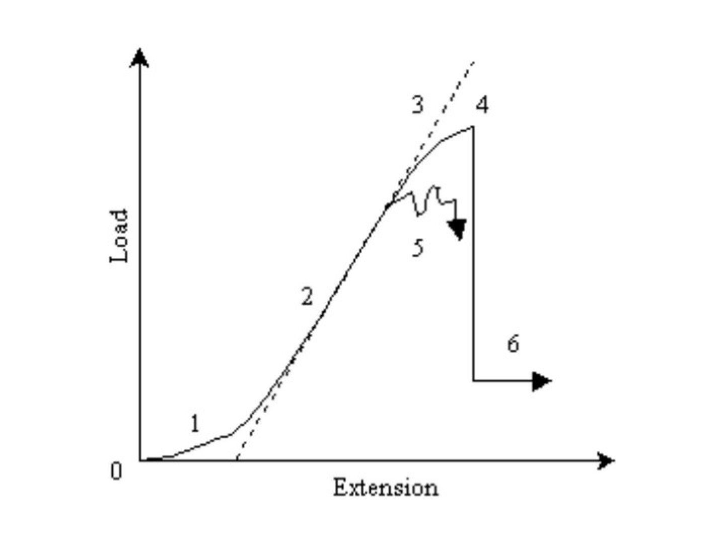
Question list
Q1-Q55 questions — tap to reveal all answerslist
- Describe the stress-strain curve of tendon and ligament.
- How do tendon and ligament stress-strain curves differ?
- Describe the stress-strain curve of ligamentum flavum.
- What is the difference between load-elongation and stress-strain curves?
- What determines the overall behaviour of ligaments and tendons on the stress-strain curve?
Answers · Q & A
Q1.Describe the stress-strain curve of tendon and ligament.
- Toe region - crimp; Modulas of elasticity is not constant, ligament becomes stiffer as more collagen fibrils are recruited (ends about 2% strain)
- Linear region - all fibres straightened; intermolecular sliding of collagen triple helices (<4% strain)
- Yield point and ultimate tensile strength
- Progressive failure (4-8% strain): sequential fibril failure, irreversible plastic deformation
- Complete rupture (>8% strain)
Q2.How do tendon and ligament stress-strain curves differ?
- Toe region is much less prominent in tendon as fibres are more aligned
- Linear region is similar
- Ultimate tensile strength is higher in tendon
Q3.Describe the stress-strain curve of ligamentum flavum.
- High elastin content
- Tolerates high strain up to 50% before stiffness increases
- Stiffness then rises significantly with further loads before it fails abruptly
Q4.What is the difference between load-elongation and stress-strain curves?
- Stress-strain curve is normalised with respect to specimen dimensions
- Load-elongation curve depends on the size of the specimen
Q5.What determines the overall behaviour of ligaments and tendons on the stress-strain curve?
- The individual crimp structure of the collagen fibres
- The sequential failure of collagen fibrils along the curve