▸ Slide 14 · Bone and jointBone and Joint · 2 questions expand
Q1-Q22 questions — tap to reveal all answerslist
- What is the theme of this section?
- List the bone and joint topics covered in this section.
Q1.What is the theme of this section?
- Bone and joint
- Title of this section divider slide
Q2.List the bone and joint topics covered in this section.
- Not covered in the speaker notes
- Topics are covered individually on later slides
▸ Slide 15 · Tell me about boneBone and Joint · 11 questions expand
Q1-Q1111 questions — tap to reveal all answerslist
- What is bone?
- What are the functions of bone?
- What types of bone are seen macroscopically and where are they found?
- What are the microscopic components of bone?
- What are the organic and inorganic components of bone ECM?
- What mnemonic summarises the lamellar vs woven bone differences?
- Differentiate lamellar and woven bone.
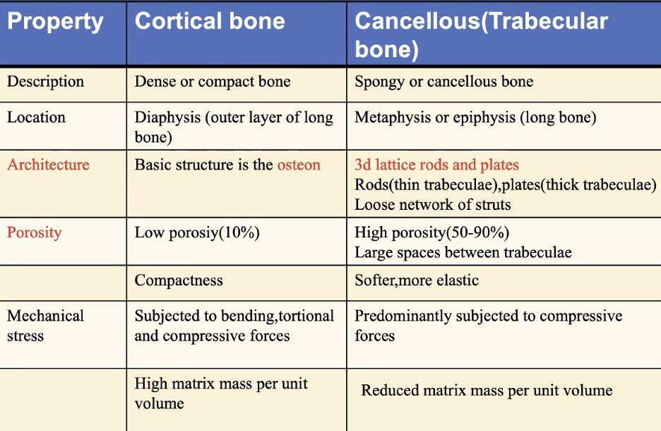
- Differentiate cortical and cancellous bone.
- Describe the structure of the Haversian system.
- What is the role of Volkman canals?
- What is Wolff's law?
Q1.What is bone?
- A solid organ
- A specialised connective tissue with several main functions
Q2.What are the functions of bone?
- Haematopoiesis
- Calcium reservoir
- Skeletal framework for locomotion
Q3.What types of bone are seen macroscopically and where are they found?
- Lamellar bone (mature) is divided into cortical and cancellous bone
- Woven bone (primitive, immature) is found in fracture callus and distraction histiogenesis
Q4.What are the microscopic components of bone?
- Comprises cells (10%) and ECM (90%)
- Cells: osteoblasts, osteoclasts, osteocytes, bone lining cells, osteoprogenitor cells
Q5.What are the organic and inorganic components of bone ECM?
- Organic 40%: type 1 collagen, proteoglycans, matrix proteins (osteocalcin, osteonectin, osteopontin), growth factors and cytokines
- Inorganic 60%: HA (hydroxyapatite)
Q6.What mnemonic summarises the lamellar vs woven bone differences?
- DLC OBT – Daniel's little cafe offers best tea
- Description, Location, Composition, Organisation, Biomechanics, Turnover
Q7.Differentiate lamellar and woven bone.
- Lamellar: mature, less cellular, ordered stress-oriented collagen, anisotropic, more stiff/less ductile, low turnover
- Lamellar is formed by enchondral or intramembranous ossification
- Woven: primitive/immature, more cellular, dense collagen fibres, no lamellae, isotropic, less stiff/more ductile, high turnover
Q8.Differentiate cortical and cancellous bone.
- Cortical (compact): diaphysis, osteon/Haversian system along the longitudinal axis, low porosity, higher Young's modulus, low turnover
- Cancellous (spongy): metaphysis, 3D lattice of trabeculae, porosity 50-90%, lower Young's modulus, compressive forces, high turnover
Q9.Describe the structure of the Haversian system.
- Central neurovascular channel enclosed within concentric lamellae
- Each osteon has 5-7 concentric rings; collagen arranged in a herringbone pattern
- Separated by a cement line where canniculi and collagen do not cross, an area of relative weakness and fracture propagation
- Interstitial lamellae lie between osteons
Q10.What is the role of Volkman canals?
- Run perpendicular to the long bone axis
- A low pressure system
- Make blood flow in the long bone centrifugal (in to out)
Q11.What is Wolff's law?
- Bone models and remodels in response to the mechanical stresses it experiences
- resulting in a minimal weight structure that is adapted to its applied stresses
▸ Slide 16 · Haematopietic monocyte/ granulocyte cell lineageBone and Joint · 7 questions expand
Q1-Q77 questions — tap to reveal all answerslist
- From which lineage do osteoclasts arise?
- What are osteoclasts and how are they activated?
- Which enzymes do osteoclasts use to hydrolyse organic matrix?
- List the steps of osteoclast bone resorption.
- What activates osteoclasts? (PR6)
- What inhibits osteoclasts? (CEO)10
- Name other multinucleated giant cells.
Q1.From which lineage do osteoclasts arise?
- The haematopoietic monocyte/granulocyte cell lineage
Q2.What are osteoclasts and how are they activated?
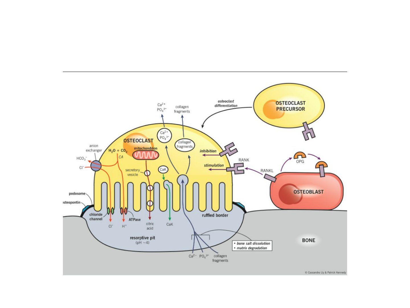
- Multinucleated giant cells responsible for bone resorption
- RANKL from osteoblasts binds immature osteoclasts, stimulating differentiation into active mature osteoclasts
Q3.Which enzymes do osteoclasts use to hydrolyse organic matrix?
- TRAP (tartrate-resistant isoenzymes of acid phosphatase)
- Cysteine proteinases e.g. cathepsins
- These are acidic proteolytic lysosomal enzymes
Q4.List the steps of osteoclast bone resorption.
- Migrate and attach to bone
- Polarisation: orientate the ruffled border to bone
- HA dissolution and matrix degradation
- Degraded bone resorbed to lacunae
- Apoptosis
Q5.What activates osteoclasts? (PR6)
- Continuous PTH acting on the osteoblast increases RANKL expression
- RANKL (NfkappaB activator ligand) from the osteoblast
- IL-6
Q6.What inhibits osteoclasts? (CEO)10
- Calcitonin
- IL-10
- Osteoprotegerin
- Estrogen (decreases RANKL)
Q7.Name other multinucleated giant cells.
- Normal: osteoclast, megakaryocyte
- Inflammation: macrophages, Kupffer cells, Langerhans cells
- Neoplasm: Hodgkin, epithelial tumour
▸ Slide 17 · OsteoblastBone and Joint · 9 questions expand

Q1-Q99 questions — tap to reveal all answerslist
- From which cells are osteoblasts derived?
- What are the characteristics of osteoblasts?
- What are the functions of osteoblasts?
- What controls osteoblast differentiation?
- What activates osteoblasts? (PED)
- What inhibits osteoblasts? (PPS)
- What are the osteotropic factors? (PPD11)
- Which embryogenesis proteins also regulate osteoblast differentiation?
- What is the role of Wnt/beta-catenin signalling in bone?
Q1.From which cells are osteoblasts derived?
- Osteoprogenitor cells, a mesenchymal stem cell line
- BMP stimulates mesenchymal cells to become osteoprogenitor cells
Q2.What are the characteristics of osteoblasts?
- Abundant organelles for synthesis: Golgi apparatus, ER, mitochondria
- Increased ALP activity and expression
Q3.What are the functions of osteoblasts?
- Bone forming: lay down osteoid (type 1 collagen + GAGs + osteocalcin, osteonectin) and are responsible for its mineralisation
- Activate osteoclasts via the RANKL system
Q4.What controls osteoblast differentiation?
- Runx2/CBFA1 is the key molecular switch in commitment of osteoprogenitors to the osteoblast lineage, with cofactor Osterix
- Osterix plays the important role later, in preosteoblast differentiation into mature osteoblasts
Q5.What activates osteoblasts? (PED)
- Pulsatile PTH
- Estrogen (inhibits adenyl cyclase, decreasing RANKL synthesis)
- 1,25 vitamin D (increases osteoblast matrix production and RANKL)
Q6.What inhibits osteoblasts? (PPS)
- Glucocorticoid
- PTH (continuous)
- PGE2 (activates adenyl cyclase)
Q7.What are the osteotropic factors? (PPD11)
- Vit D, PGE2, IL 11, PTH
- They induce osteoclast formation by upregulating RANKL
Q8.Which embryogenesis proteins also regulate osteoblast differentiation?
- HOXA10, DLX and WNT
- Typical of embryogenesis but also function in the adult skeleton to support osteoblast differentiation
Q9.What is the role of Wnt/beta-catenin signalling in bone?
- Wnt binds frizzled receptors with LRP5/6 co-receptors, stabilising beta-catenin and activating gene transcription
- Favours osteoblastic differentiation, osteoblast maturation and survival
- Inhibits osteoclast generation by increasing osteoprotegerin (OPG)
- Sclerostin (SOST gene, inhibited by RUNX2) antagonises LRP5/6
▸ Slide 18 · Osteoprogenitor cellBone and Joint · 2 questions expand
Q1-Q22 questions — tap to reveal all answerslist
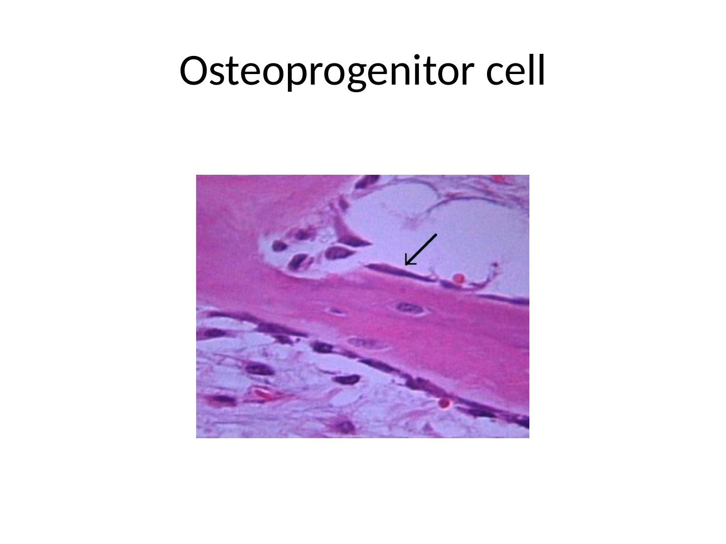
- What can osteoprogenitor cells mature into?
- Which theory describes this differentiation?
Q1.What can osteoprogenitor cells mature into?
- Osteoblast, chondroblast or fibroblast
- Depending on strain and oxygen tension
Q2.Which theory describes this differentiation?
- Perren's strain theory
- Maturation depends on strain and oxygen tension
▸ Slide 19 · OsteocyteBone and Joint · 4 questions expand

Q1-Q44 questions — tap to reveal all answerslist
- What is an osteocyte?
- What are the functions of osteocytes?
- What are the characteristics of osteocytes?
- What are bone lining cells?
Q1.What is an osteocyte?
- Described as an osteoblast trapped in osteoid
- Makes up 90% of cells in the mature skeleton
Q2.What are the functions of osteocytes?
- Regulate bone response to hormonal and mechanical stimuli
- Regulate calcium and phosphorus concentration in bone
- Stimulated by calcitonin, inhibited by PTH
Q3.What are the characteristics of osteocytes?
- High NC (minimal organelle)
- Long cytoplasmic processes
- No ALP expression
Q4.What are bone lining cells?
- Inactive osteoblasts
- Reactive for bone formation, or prepare bone for osteoclastic resorption upon PTH stimulation
▸ Slide 20 · Bone MatrixBone and Joint · 6 questions expand

Q1-Q66 questions — tap to reveal all answerslist
- What are the proportions of organic and inorganic bone matrix?
- What does the organic matrix contain and what strength does it provide?
- What are the roles of osteocalcin?
- What are the roles of osteonectin and osteopontin?
- What is the inorganic component of bone matrix?
- Which bone matrix markers can be detected in urine and what do they indicate?
Q1.What are the proportions of organic and inorganic bone matrix?
- Organic 40%
- Inorganic 60%
Q2.What does the organic matrix contain and what strength does it provide?
- Collagen type I (90%) for tensile strength; hydroxyproline forms the triple helix with glycine
- Proteoglycans for compressive strength
- Non-collagen matrix proteins: osteocalcin, osteonectin, osteopontin
- Growth factors and cytokines (TGF, IL-1, IL-6)
Q3.What are the roles of osteocalcin?
- From the mature osteoblast (marker of osteoblastic differentiation)
- Controls osteoclasts: stimulates 1,25 vitamin D, inhibits PTH
- Urine detection indicates bone maturation
Q4.What are the roles of osteonectin and osteopontin?
- Osteonectin: from osteoblast/platelets, regulates mineralisation, binds calcium
- Osteopontin (pont = bond): cell binding
Q5.What is the inorganic component of bone matrix?
- Mainly provides compressive strength
- Calcium hydroxyapatite Ca10(PO4)6(OH)2
- Osteocalcium phosphate
Q6.Which bone matrix markers can be detected in urine and what do they indicate?
- Urine hydroxyproline: osteoblast turnover marker
- Urine osteocalcin: bone maturation
▸ Slide 21 · CalciumBone and Joint · 5 questions 2 check expand

Q1-Q55 questions — tap to reveal all answerslist
- What is the distribution of calcium in the body?
- What are the functions of calcium?
- Where and how is calcium taken up?
- How is calcium excreted?
- What is the distribution and function of phosphate?
Q1.What is the distribution of calcium in the body?
- ~99% in bone
- ~1% extracellular, <1% intracellular
- Normal level 2.2-2.6 mmol/L
Q2.What are the functions of calcium?
- Neuromuscular function
- Clotting/metabolic function
- Bone mineralisation
Q3.Where and how is calcium taken up?
- Duodenum: active transport
- Jejunum: passive diffusion
Q4.How is calcium excreted?
- Kidney: 98% reabsorbed at proximal tubules (per speaker notes)
- Stool
Q5.What is the distribution and function of phosphate?
- 86% in bone, 14% intracellular, <1% plasma
- Bone mineralisation and enzyme/molecular interaction
- Uptake from food; kidney 100% resorb (per speaker notes)
Kidney reabsorbs 98% of calcium at the proximal tubules — imprecise — 98-99% of filtered calcium is reabsorbed overall; the proximal tubule accounts for only ~60-70%, with the rest reabsorbed in the loop of Henle and distal nephron — source
Kidney 100% resorbs phosphate — misleading — About 80-90% of filtered phosphate is normally reabsorbed (mostly in the proximal tubule); reabsorption approaches 100% only with very low dietary phosphate intake — source
▸ Slide 22Bone and Joint · 4 questions expand
Q1-Q44 questions — tap to reveal all answerslist
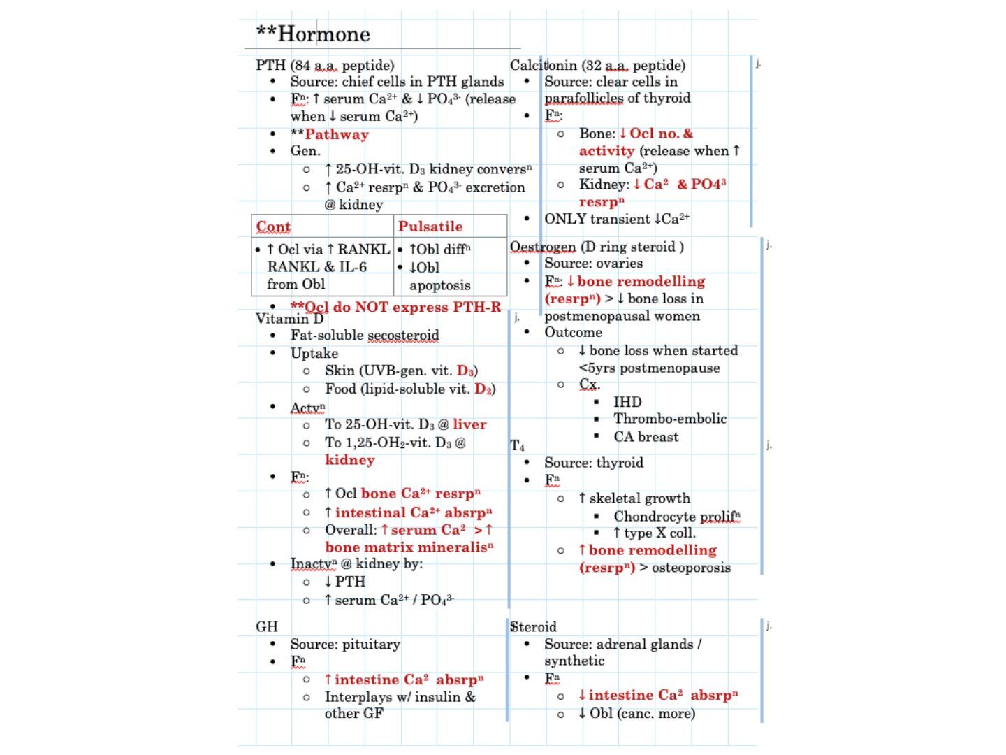
- How do PTH and calcitonin differ in their effects on calcium homeostasis?
- What is the difference between pulsatile and continuous PTH action on bone?
- Describe the activation and actions of vitamin D.
- What are the skeletal effects of estrogen and thyroxine (T4)?
Q1.How do PTH and calcitonin differ in their effects on calcium homeostasis?
- PTH (parathyroid chief cells): raises serum Ca2+, lowers PO4^3-; released when serum Ca2+ falls
- Calcitonin (thyroid parafollicular cells): lowers osteoclast number/activity and renal Ca2+/PO4^3- resorption; released when serum Ca2+ rises
- Calcitonin causes only a transient fall in Ca2+
Q2.What is the difference between pulsatile and continuous PTH action on bone?
- Pulsatile PTH: increases osteoblast differentiation and decreases osteoblast apoptosis
- Continuous PTH: increases osteoclast activity via RANKL and IL-6 from osteoblasts
- Osteoclasts do not express PTH receptors
Q3.Describe the activation and actions of vitamin D.
- Fat-soluble secosteroid; sources are skin (UVB-generated vitamin D3) and food
- Activated to 25-OH-vit D3 in the liver, then to 1,25-OH2-vit D3 in the kidney
- Increases intestinal Ca2+ absorption and osteoclastic bone resorption, raising serum Ca2+
Q4.What are the skeletal effects of estrogen and thyroxine (T4)?
- Estrogen (ovarian D-ring steroid): decreases bone remodelling and postmenopausal bone loss; benefit when started <5 years postmenopause
- Estrogen risks: IHD, thrombo-embolic events and breast cancer
- T4 (thyroid): increases skeletal growth and bone remodelling, leading to osteoporosis
▸ Slide 23 · Cutting coneBone and Joint · 5 questions expand
Q1-Q55 questions — tap to reveal all answerslist
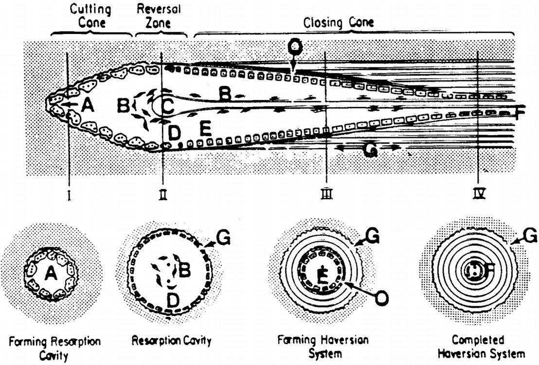
- What is the sequence of cellular events in a cutting cone?
- What are the labelled components of a cutting cone?
- How does the cutting cone tunnel change as it advances?
- From which direction do osteoblasts deposit new bone in a cutting cone?
- Which cells line the haversian canal after the cutting cone has passed?
Q1.What is the sequence of cellular events in a cutting cone?
- Osteoclasts at the front to remove bone
- Followed by a layer of osteoblasts depositing successive centripetal layers of lamellae and osteoid
- Tunnel size narrows to a haversian canal
Q2.What are the labelled components of a cutting cone?
- A osteoclast
- B osteoprogenitor cells
- C capillary
- D mononuclear cell lining reversal zone
- E osteoblast apposing bone centripetally
- F flattened osteoblast/osteoclast lining haversian canal
- G osteoid
Q3.How does the cutting cone tunnel change as it advances?
- Tunnel size narrows
- Eventually forms the haversian canal
Q4.From which direction do osteoblasts deposit new bone in a cutting cone?
- Centripetally (towards the centre of the tunnel)
- Deposit successive layers of lamellae and osteoid
- Following behind the osteoclast front
Q5.Which cells line the haversian canal after the cutting cone has passed?
- Flattened osteoblast/osteoclast lining the haversian canal
- Mononuclear cells lining the reversal zone
▸ Slide 24 · Draw a structure of haversian systemBone and Joint · 4 questions expand

Q1-Q44 questions — tap to reveal all answerslist
- What is the haversian system (osteon)?
- What is the role of Volkmann canals?
- What are osteocytes, lacunae and canaliculi?
- What lamellar types are described in cortical bone?
Q1.What is the haversian system (osteon)?
- Basic structural unit of cortical bone
- Lies parallel to the long axis of bone
- Central haversian canal transmits a neurovascular bundle
- Concentric lamellae of collagen arranged in a herringbone pattern
- Cement lines separate the osteons
Q2.What is the role of Volkmann canals?
- Run transversely to the bone long axis
- Permit communication between outer periosteal vessels and the haversian canal
Q3.What are osteocytes, lacunae and canaliculi?
- Osteocytes are trapped osteoblasts located within lacunae between lamellae
- Canaliculi are canals occupied by osteocyte cell processes connecting lacunae
- Modulate the response of bone to mechanical stimuli
- Allow exchange of nutrients and waste products to maintain osteocyte viability
Q4.What lamellar types are described in cortical bone?
- Concentric lamellae
- Interstitial lamellae
- Separated by cement lines
▸ Slide 25 · Describe the structure of periosteumBone and Joint · 3 questions expand

Q1-Q33 questions — tap to reveal all answerslist
- Describe the structure of periosteum.
- What are the functions of periosteum?
- Which periosteal layer is osteogenic and why?
Q1.Describe the structure of periosteum.
- Outer layer of fibroblasts
- Inner layer of osteoblasts (cambial layer)
- Outer layer: structural support, less cellular, continuous with joint capsule
- Inner cambial layer: vascular, osteogenic, contributes to bone growth and fracture healing
Q2.What are the functions of periosteum?
- Medium through which tendon, muscles and ligaments attach
- Nutrition
- Forms bone when required
- Protective membrane for bone tissue
Q3.Which periosteal layer is osteogenic and why?
- Inner cambial layer
- Vascular and contributes to bone growth and fracture healing
▸ Slide 26 · Tell me about bone healingBone and Joint · 9 questions expand
Q1-Q99 questions — tap to reveal all answerslist
- What is Perren's strain theory?
- Describe primary bone healing.
- Describe the stages of secondary bone healing.
- What adjuncts aid fracture healing?
- Differentiate bone modelling from remodelling.
- What is the role of fibrinolysis in fracture healing?
- Describe enchondral ossification during soft callus formation.
- Where does intramembranous ossification occur in secondary bone healing?
- What is the bone healing nonunion (BHN) model?
Q1.What is Perren's strain theory?
- The degree of interfragmentary strain governs the cellular response and therefore the tissue that forms
- <2% = primary bone healing
- 2-10% = secondary bone healing
- >10% = nonunion / fibrous union
Q2.Describe primary bone healing.
- It is actually bone remodelling - haversian cutting cones cross the fracture site and directly lay down osteons
- It is a process of intramembranous ossification where there is no intermediate step of cartilage formation, therefore no callus formation
- Contact healing: direct bone apposition with gap <0.01mm
- Gap healing: gap <1mm; vessels grow into the gap and osteoblasts lay lamellar or woven bone
- Followed by haviersian remodelling by cutting cones (osteoclast reams, vessel grows in, trailing osteoblasts lay new bone)
Q3.Describe the stages of secondary bone healing.
- Hematoma and inflammation (<1wk): haematoma brings in haemtopoetic cells. Inflammatory mediators lead to granulation and angiogenesis; osteoclasts and macrophages remove debris
- Soft callus (1 weeks to 1 month): endochondral ossification at intermediate strain/low O2; intramembranous ossification at low strain/high O2
- Hard callus (1-4 months): soft calcified chondral callus remodels into hard mineralised osteoid callus
- Remodelling: Wolff's law and piezoelectric charges dictate distribution of remodelling
Q4.What adjuncts aid fracture healing?
- Low intensity pulsed USG increases nanomotion across the fracture site, increasing healing and callus strength
- Bone stimulator (electromagnetic stimulation) increases BMP concentration and osteoblastic differentiation
Q5.Differentiate bone modelling from remodelling.
- Modelling: change of shape or growth by independent, simultaneous actions of osteoblast and osteoclast
- Remodelling: replacement of the same amount of matrix, sequential osteoblast then osteoclast within the same BMU
- Cancellous remodelling: osteoclastic resorption + osteoblastic deposition of lamellar bone
- Cortical remodelling: cutting cone
Q6.What is the role of fibrinolysis in fracture healing?
- Plays a key transition step from haematoma formation to angiogenesis and fracture healing
- Cited from NEJM 2015 O'Keiffe
Q7.Describe enchondral ossification during soft callus formation.
- Occurs in areas of intermediate strain and low O2 tension
- Stem cells differentiate into chondrocytes, laying down type I collagen and fibrocartilaginous matrix
- Followed by vascularisation and osteoblasts laying down bone on the cartilaginous scaffold
- Forms the interfragmentary callus and external callus
Q8.Where does intramembranous ossification occur in secondary bone healing?
- At sites of low strain and high O2 tension (when fracture strain decreases)
- Osteoblasts from the cambrium layer of periosteum directly lay down woven bone
- Woven bone is later remodelled to lamellar bone
- Forms the medullary callus and periosteal callus
Q9.What is the bone healing nonunion (BHN) model?
- Described by Elliot, JBJS 2016
- Unifies Perren's strain theory, Wolff's law and Frost's concept of mechanostat
- Tissue around a fracture is called a bone healing unit, producing a physiological response to its biological and mechanical environment
- The lecturer suggests drawing the graph
▸ Slide 27Bone and Joint · 2 questions expand

Q1-Q22 questions — tap to reveal all answerslist
- What bone and joint topic is shown on this slide?
- What are the key learning points from this slide?
Q1.What bone and joint topic is shown on this slide?
- Not covered in the speaker notes - the slide image is the only source
Q2.What are the key learning points from this slide?
- Not covered in the speaker notes
▸ Slide 28 · Xray showing features of renal osteodystrophyBone and Joint · 6 questions expand

Q1-Q66 questions — tap to reveal all answerslist
- What are the X-ray features of renal osteodystrophy?
- How is renal osteodystrophy classified and what causes the bone mineralisation deficiency?
- What causes low-turnover renal osteodystrophy?
- How does renal osteodystrophy present?
- What is the management of renal osteodystrophy?
- Describe the pathophysiology of high-turnover renal osteodystrophy.
Q1.What are the X-ray features of renal osteodystrophy?
- Subperiosteal resorption
- Rugger jersey spine
- Brown tumour
- Tumoral calcinosis (lecturer advises avoiding this term)
- All are features of high bone turnover disease
Q2.How is renal osteodystrophy classified and what causes the bone mineralisation deficiency?
- High turnover disease with elevated PTH vs low turnover disease with normal PTH
- Bone mineralisation deficiency due to electrolyte and endocrine abnormalities
- X-ray features of both osteomalacia (impaired mineralisation) and hyperparathyroidism
Q3.What causes low-turnover renal osteodystrophy?
- Aluminium accumulation in renal failure
- Excess deposition into bone -> toxic to osteoblasts (differentiation and proliferation)
- Also impairs PTH release
- PTH is normal
Q4.How does renal osteodystrophy present?
- Bony problem: pathological fracture
- Soft tissue: tendinitis, tendon rupture, carpal tunnel syndrome
- Growth retardation, deformity, SCFE in children
Q5.What is the management of renal osteodystrophy?
- Treat the underlying cause, multidisciplinary approach
- Aluminium chelator
- Calcium supplement
- Vitamin D supplement
- Bisphosphonates
Q6.Describe the pathophysiology of high-turnover renal osteodystrophy.
- Uraemia -> abnormal PO4 excretion -> PO4 retention (glomerular) + kidney unable to convert vitamin D to active form (tubules) -> hypocalcaemia
- Failure of 1-alpha hydroxylase -> hypocalcaemia
- High PO4 and low Ca -> increased PTH -> hyperplasia of chief cells of the parathyroid gland
▸ Slide 29 · Type of jointBone and Joint · 5 questions expand
Q1-Q55 questions — tap to reveal all answerslist
- How are joints classified?
- Classify joints by nature with examples.
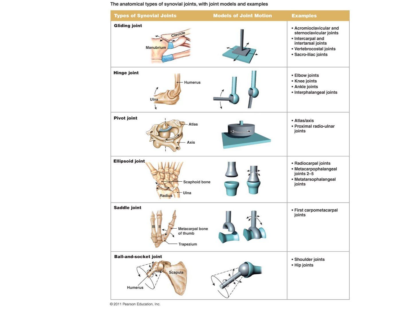
- Which joints are given as examples of the 6 morphological types of synovial joint?
- Classify joints by movement.
- How many morphological types of synovial joint are there?
Q1.How are joints classified?
- By anatomical composition
- By movement allowed
Q2.Classify joints by nature with examples.
- Synovial
- Cartilaginous: symphysis, synchondrosis, IVD
- Fibrous: syndesmosis, suture
Q3.Which joints are given as examples of the 6 morphological types of synovial joint?
- C1/2
- SCJ
- GHJ
- Elbow, radiocarpal, 1st CMCJ
Q4.Classify joints by movement.
- Diarthrosis
- Amphiarthrosis (pubic symphysis)
- Synarthrosis (suture)
Q5.How many morphological types of synovial joint are there?
- 6 types of synovial joint, classified by morphology
▸ Slide 30 · Bone graftBone and Joint · 15 questions expand
Q1-Q1515 questions — tap to reveal all answerslist
- Define bone graft and the properties of an ideal graft.
- What are the common uses of bone graft?
- How is a cancellous bone graft incorporated?
- How is a cortical bone graft incorporated?
- Compare autograft with allograft.
- What are the methods of allograft preservation?
- How are bone grafts classified?
- By what mechanisms are bone grafts incorporated into host bone?
- What factors affect bone graft uptake?
- What are the non-vascularised sources of autograft?
- Describe the ASIS autograft harvest and the structure at risk.
- Describe the PSIS autograft harvest and the structures at risk.
- What are the vascularised sources of autograft?
- How is a bone bank set up and allograft processed?
- What is the principle of graft fixation - nail vs plate?
Q1.Define bone graft and the properties of an ideal graft.
- Implanted materials to promote bone healing
- Biological properties: osteoconduction, osteoinduction, osteogenesis, vascularity
- Structural support
Q2.What are the common uses of bone graft?
- AJR - structural support (bone defect management)
- Spine - induce fusion
- Trauma - fracture nonunion
Q3.How is a cancellous bone graft incorporated?
- Inflammation phase: vascular ingrowth, chemotaxis
- Revascularisation phase (0-3 weeks)
- Creep substitution (3-12 weeks): angiogenesis, osteoblasts and osteoclasts introduced and work at the same time
- Remodelling phase (>12 weeks)
- Incorporation can be complete; no osteopenia during incorporation
Q4.How is a cortical bone graft incorporated?
- Initial incorporation at the host-graft junction by endochondral bone formation
- Sequential action of osteoclast then osteoblast: graft bone removed by cutting cones then new bone laid down
- 50% loss of strength in 3-6 months, then fully returns over 1-2 years
- Incorporation is not complete; initial osteopenia on X-ray during bone absorption
- Called reverse creep substitution
Q5.Compare autograft with allograft.
- Autograft pros: osteogenesis, no immunogenicity, less chance of infection
- Autograft cons: limited supply, donor site morbidity
- Allograft requires donor consent, screening, processing and storage
- Aim of bone bank processing: decrease immune sensitisation and disease transmission
- Other sources include synthetic bone substitute and xenograft
Q6.What are the methods of allograft preservation?
- Fresh frozen at -70deg: preserves BMP, better mechanical strength; immunogenic, disease transmission risk; shelf life 2 years at -20deg, 5 years at -70deg
- Freeze dry (lyophilise): less immunogenic, less disease transmission, longer shelf life; weak structure, no BMP - purely osteoconductive
- Fresh
Q7.How are bone grafts classified?
- By source: autograft / allograft / synthetic / xenograft
- By structure: cortical or cancellous
Q8.By what mechanisms are bone grafts incorporated into host bone?
- Inflammation
- Osteoblast differentiation
- Osteoinduction and osteoconduction
- Remodelling
Q9.What factors affect bone graft uptake?
- Biological: local vs systemic
- Mechanical
- Think of the factors affecting fracture healing
Q10.What are the non-vascularised sources of autograft?
- Femoral head
- ASIS
- PSIS
Q11.Describe the ASIS autograft harvest and the structure at risk.
- 2cm posterior to ASIS, 3cm wound along the crest
- Subperiosteal elevation of iliacus
- Lateral cutaneous nerve of thigh is at risk
Q12.Describe the PSIS autograft harvest and the structures at risk.
- 1cm lateral to PSIS, longitudinal incision
- Subperiosteal elevation of gluteal muscle
- Superior cluneal nerve L1-3 (6cm lateral to PSIS) at risk
- Superior gluteal NV (6cm inferior to PSIS) at risk
Q13.What are the vascularised sources of autograft?
- Wrist: 1,2 intercompartmental supraretinacular (ICSR) bone graft for scaphoid; 4,5 ICSR bone graft for lunate AVN
- Fibular
- Iliac crest (deep iliac circumflex artery)
Q14.How is a bone bank set up and allograft processed?
- Donor consent and screening (IVDU, blood for HIV, Hep B/C, syphilis, Rhesus status)
- Exclusion criteria: HIV, Hep B/C, malignancy, RA/autoimmune disease, steroid
- Processing: physical debridement, ultrasonic processing (remove cells/blood), ethanol (denature protein), antibiotic soak, irradiation, +/- demineralisation
- Then storage
Q15.What is the principle of graft fixation - nail vs plate?
- Nail > plate
- Even stress distribution
- Increased time for incorporation, needs a rigid device
- Increase contact area
- Never remove the implant
▸ Slide 31 · Synthetic:Bone and Joint · 7 questions 2 check expand

Q1-Q77 questions — tap to reveal all answerslist
- What are the synthetic bone graft types?
- Describe tricalcium phosphate.
- What does DBM contain and not contain?
- How do bone morphogenic proteins work?
- What are the contraindications to BMP?
- What are the complications of BMP?
- How is DBM manufactured and what does the acidic extraction leave behind?
Q1.What are the synthetic bone graft types?
- Osteoconduction: tricalcium phosphate
- Osteoinduction: bone morphogenic protein 2 and 7
- Others: demineralised bone matrix, hydroxyapatite, calcium phosphate, calcium sulphate
Q2.Describe tricalcium phosphate.
- Ceramic scaffold providing a scaffold for ingrowth of vessels and osteogenic cells
- Porosity 50% - balance between mechanical strength and porosity
- Macropore 100-500mcg for ingrowth of vessels and cells
- Micropore 10mcg increases surface area of remodelling
- Available as blocks, wedges and granules; trade name Chronos (Synthes)
Q3.What does DBM contain and not contain?
- Contains: collagen, BMP, transforming growth factor-beta, residual calcium
- Does NOT contain mesenchymal precursor cells
- Made by acidic extraction of bone matrix from allograft, then sterilised (may decrease BMP availability)
- Osteoconductive without structural support; minimally osteoinductive
- Interproduct and interlot variability common
Q4.How do bone morphogenic proteins work?
- Osteoinductive; belong to the TGF-beta superfamily
- Stimulate undifferentiated perivascular mesenchymal cells to differentiate into osteoblasts via serine-threonine kinase receptors
- Activate intracellular signalling molecules called SMAD
- rhBMP-2 and rhBMP-7 are FDA-approved for application in long bones and spine
- BMP-2 marketed by Medtronic; BMP-7 by Stryker
Q5.What are the contraindications to BMP?
- Pregnancy
- Allergy to bovine type I collagen
- Infection
- Tumor
- Skeletal immaturity
Q6.What are the complications of BMP?
- Under- or overproduction of bone (heterotopic ossification)
- Inflammatory responses / seroma formation
- Not for cervical spine - increased cervical swelling
- Early bone resorption
Q7.How is DBM manufactured and what does the acidic extraction leave behind?
- Acidic extraction of bone matrix from allograft removes the minerals
- Leaves collagen, non-collagenous proteins and bone growth factors
- BMP quantity is extremely low and variable
- Sterilisation may decrease the availability of BMP
rhBMP-2 and rhBMP-7 are FDA-approved for application in long bones and spine — outdated / imprecise — rhBMP-2 (INFUSE) is approved for anterior lumbar interbody fusion and acute open tibial shaft fractures; rhBMP-7 (OP-1) was approved via HDE only for recalcitrant long-bone nonunion and is no longer marketed in the US — source
Macropore 100-500mcg and micropore 10mcg increase remodelling surface area — unit error — pore sizes are measured in micrometres (um), not micrograms: macropores ~100-500um and micropores ~10um or less — source